
Before you read: At Skinpaths, we distinguish between opinion and evidence. This content is based on indexed medical literature and recommendations from scientific societies. At the end, you will find a complete bibliography for verification and further study.
Learning objectives
-
Define dermoepidermal junction (DEJ) and basement membrane zone (BMZ) in normal skin.
-
Recognizing the UDE in H&E and knowing when to add PAS .
-
Explain the ultrastructure (hemidesmosome–lamina lucida–lamina densa–sublamina densa) and its histological “translation”.
-
Locate key proteins (integrins, laminins, collagen IV, collagen VII, plectin/BP230) on a functional mental map.
-
Describe normal variations (anatomical site/age) of the dermis-epidermis interface.
Founding facts (what you absolutely must know)
-
The epidermal dermal junction (EDJ) is an adhesive and signaling interface zone that joins the epidermis to the dermis and organizes cellular niches. Breitkreutz 2013
-
“MB” or “BMZ” is a molecular/ultrastructural term; in H&E staining it is often subtle or not visible as a “line”. Breitkreutz 2013
-
The basement membrane is composed of networks of laminins and collagen IV , connected by proteoglycans (e.g., perlecan) and other molecules. Behrens 2012 Lennon 2025
-
Collagen IV predominates in the lamina densa (basal scaffolding). Abreu-Velez 2012
-
Laminin-332 is a major component of the UDE and constitutes a functional “bridge” between the basal keratinocyte and the matrix. Kiritsi 2012
-
The hemidesmosome is a basal “rivet”: integrin α6β4, collagen XVII (BP180), plectin, and BP230 connect membrane and keratins. Walko 2014
-
The typical basal cytoskeleton is dominated by 5/14 keratins , anchored to hemidesmosomes via plectin. Bouameur 2014
-
Collagen VII forms anchoring fibrils in the sublamina densa, connecting the basement membrane to the papillary dermis. Chung 2010
-
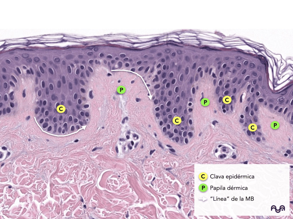
Rete ridges (epidermal clubs) and dermal papillae increase the contact surface between the two and provide mechanical resistance (interlock). Aleemardani 2021
-
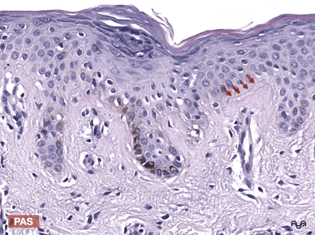
In PAS staining, the basement membrane is often highlighted by its carbohydrate-rich components (glycoproteins/proteoglycans). This is useful when H&E does not clearly show the interface. Breitkreutz 2013
-
The “lamina lucida/densa” (not to be confused with the stratum lucidum of the epidermis in glabrous skin) is a concept of electron microscopy ; (in conventional microscopy) it is inferred by special stains and by tissue organization. Breitkreutz 2013
-
Normal variation: in thick skin (palms/soles) the interface is usually more wavy (more pronounced ridges). Aleemardani 2021
-
With age and depending on the anatomical location, the joint architecture can change (e.g., relative flattening), with its consequent biomechanical impact. Aleemardani 2021
Key concepts
1) Useful Definitions
-
Dermoepidermal junction (DEJ): the “territory” where the epidermis and dermis interdigitate (ridges and papillae) and are mechanically and biologically coupled. Aleemardani 2021
-
Basement membrane zone (BMZ): the set of proteins and networks that form the basement membrane and its anchoring complexes (includes hemidesmosomes and underlying structures). Breitkreutz 2013 Lennon 2025
2) Functions in normal skin
-
Adhesion and resistance to mechanical stress (preventing the epidermis from «sliding» over the dermis). Walko 2014
-
Barrier and compartmentalization (organizes microenvironments between epithelium and stroma). Breitkreutz 2013
-
Outside-in signaling via integrins/laminins that influences survival, migration, and differentiation. Kiritsi 2012
Histological presentation of the UDE
A) Light microscopy (H&E): what to look for
-
Epidermal basal layer : row of basal keratinocytes on the boundary with the dermis; more basophilic nuclei, sometimes with a subtle «palisade» arrangement depending on the section.
-
Epidermal ridges/clavas (rete ridges) : extensions of the epidermis into the dermis.
-
Dermal papillae : projections of the papillary dermis into the epidermis (increasing surface area). Aleemardani 2021
-
Basement membrane “line” : in H&E staining it may be faint; do not assume it is “not there” if you do not see it clearly (limitation of the technique). Breitkreutz 2013
Practical tip: If your goal is to study the interface , select fields where there are well-oriented papillae and ridges (perpendicular section). A tangential section can artificially «flatten» the architecture.
B) PAS staining: when and why it helps
-
When: suspicion of subtle interface in H&E, teaching need to highlight contour, or comparative evaluation between areas.
-
Why: The basement membrane contains glycoproteins/proteoglycans that typically stain with PAS, outlining a more prominent band/contour. Breitkreutz 2013
The UDE at the Ultrastructural level
In electron microscopy, a classic “stack” is described:
Basal keratinocyte (5/14 keratin cytoskeleton)
→ Hemidesmosomal plaque (plectin, BP230)
→ Basal plasma membrane (integrin α6β4, collagen XVII/BP180)
→ Lamina lucida (interface zone rich in adhesion components; EM concept)
→ Dense lamina (dominant network of collagen IV + organized laminins)
→ Sublamina densa (anchoring fibrils of collagen VII towards papillary dermis)
What to memorize (the essentials):
-
Hemidesmosome = basal anchor to keratins. Walko 2014
-
Lamina densa = collagen IV as the main framework. Abreu-Velez 2012
-
Sublamina densa = collagen VII (anchoring fibrils). Chung 2010
-
Laminins (e.g., laminin-332) connect cellular networks and receptors (integrins). Kiritsi 2012 Hohenester 2013
Microscopic evaluation
Step by step to study the UDE on a normal sheet
-
Cutting orientation: look for areas perpendicular to the surface to see ridges/papillae.
-
H&E first: identify basal layer and papillary dermis; assess undulation (rete ridges).
-
If you need to outline MB: add PAS (or check if the lab has PAS available in the teaching set).
-
If your goal is to “map molecules” (teaching/research): selected immunostainings (see below).
Pitfalls (when the student gets “lost”)
-
Confusing UDE (architecture) with BMZ (molecular) : they do not always coincide as a single «visible line». Breitkreutz 2013
-
Overinterpreting a tangential cut as a “planar joint” (orientation artifact).
-
Forgetting that skin varies by anatomical site (e.g., thick vs. thin skin). Aleemardani 2021
Additional studies / markers (for normal histology)
Stains and what they highlight
-
H&E: general architecture (epidermal layers, papillary/reticular dermis, interdigitation).
-
PAS: helps to delineate the basement membrane due to its composition rich in associated carbohydrates. Breitkreutz 2013
Immunomarkers (for teaching purposes)
-
Collagen IV: traces the network of lamina densa (basement membrane). Abreu-Velez 2012
-
Laminin-332: major component of DEJ and adhesive bridge. Kiritsi 2012
-
Collagen VII: marks the sublamina densa anchoring system (fibrils). Chung 2010
-
Integrin β4 / plectin: locate hemidesmosomes/anchoring plaque in the basal lamina. Walko 2014 Schaapveld 1998
Electron microscopy
-
Useful for directly visualizing lamina lucida/densa and hemidesmosomal plaques; its use is primarily academic/research-oriented. Walko 2014
Recommended readings
Essentials
-
Epidermal basement membrane: composition and functions. — Breitkreutz 2013
-
Architecture and function of the hemidesmosome. — Walko 2014
-
Supramolecular organization: laminin vs collagen IV networks and the role of perlecan.— Behrens 2012
-
Collagen IV in normal skin (useful approach for BM).— Abreu-Velez 2012
-
Collagen VII and anchoring fibrils.— Chung 2010
To delve deeper
-
A modern view of basement membranes: composition, assembly, and function.— Lennon 2025
-
Laminin assembly in basement membranes (conceptual and powerful).— Hohenester 2013
-
Laminin-332 as an essential component of DEJ (provides the molecular “bridge”).— Kiritsi 2012
-
β4 integrin and hemidesmosomal component assembly/localization (experimental, instructional).— Schaapveld 1998
-
Desmosomes and IF (useful for contrast with hemidesmosome).— Hatzfeld 2017
-
Plectin and K5/K14 anchoring to hemidesmosomes (cytoskeleton-UDE bridge).— Bouameur 2014
-
DEJ and role of ridges/papillae (interface as a functional unit).— Aleemardani 2021
Bibliography and sources
-
Breitkreutz D, Koxholt I, Thiemann K, Nischt R. Skin basement membrane: the foundation of epidermal integrity—BM functions and diverse roles of bridging molecules nidogen and perlecan. Biomed Res Int. 2013;2013:179784. doi:10.1155/2013/179784. [show_150ms_ease-in] » data-testid=»webpage-citation-pill»> PubMed
-
Behrens DT, Villone D, Koch M, Brunner G, Sorokin L, Robenek H, et al. The epidermal basement membrane is a composite of separate laminin- or collagen IV-containing networks connected by aggregated perlecan, but not by nidogens. J Biol Chem. 2012 May 25;287(22):18700-18709. doi:10.1074/jbc.M111.336073. [show_150ms_ease-in] » data-testid=»webpage-citation-pill»> PubMed
-
Walko G, Castañón MJ, Wiche G. Molecular architecture and function of the hemidesmosome. Cell Tissue Res. 2015 Jun;360(3):529-544. doi:10.1007/s00441-015-2216-6. [show_150ms_ease-in] » data-testid=»webpage-citation-pill»> PubMed
-
Lennon R, Sherwood DR. Basement membranes at a glance. J Cell Sci. 2025 Sep 1;138(17):jcs263947. doi:10.1242/jcs.263947. [show_150ms_ease-in] » data-testid=»webpage-citation-pill»> PubMed
-
Hohenester E, Yurchenco PD. Laminins in basement membrane assembly. Cell Adh Migr. 2013 Jan-Feb;7(1):56-63. doi:10.4161/cam.21831. [show_150ms_ease-in] » data-testid=»webpage-citation-pill»> PubMed
-
Abreu-Velez AM, Howard MS. Collagen IV in Normal Skin and in Pathological Processes. N Am J Med Sci. 2012 Jan;4(1):1-8. doi:10.4103/1947-2714.92892. [show_150ms_ease-in] » data-testid=»webpage-citation-pill»> PubMed
-
Chung HJ, Uitto J. Type VII collagen: the anchoring fibril protein at fault in dystrophic epidermolysis bullosa. Dermatol Clin. 2010 Jan;28(1):93-105. doi:10.1016/j.det.2009.10.011. [show_150ms_ease-in] » data-testid=»webpage-citation-pill»> PubMed
-
Kiritsi D, Has C, Bruckner-Tuderman L. Laminin 332 in junctional epidermolysis bullosa. Cell Adh Migr. 2013 Jan-Feb;7(1):135-141. doi:10.4161/cam.22418. [show_150ms_ease-in] » data-testid=»webpage-citation-pill»> PubMed
-
Schaapveld RQ, Borradori L, Geerts D, van Leusden MR, Kuikman I, Nievers MG, et al. Hemidesmosome formation is initiated by the beta4 integrin subunit, requires complex formation of beta4 and HD1/plectin, and involves a direct interaction between beta4 and the bullous pemphigoid antigen 180. J Cell Biol. 1998 Jul 13;142(1):271-284. [show_150ms_ease-in] » data-testid=»webpage-citation-pill»> PubMed
-
Bouameur JE, Favre B, Fontao L, Lingasamy P, Begré N, Borradori L. Interaction of plectin with keratins 5 and 14: dependence on several plectin domains and keratin quaternary structure. J Invest Dermatol. 2014 Nov;134(11):2776-2783. doi:10.1038/jid.2014.255. [show_150ms_ease-in] » data-testid=»webpage-citation-pill»> PubMed
-
Aleemardani M, Trikić MZ, Green NH, Claeyssens F. The Importance of Mimicking Dermal-Epidermal Junction for Skin Tissue Engineering: A Review. Bioengineering (Basel). 2021 Oct 20;8(11):148. doi:10.3390/bioengineering8110148. [show_150ms_ease-in] » data-testid=»webpage-citation-pill»> PubMed
-
Hatzfeld M, Keil R, Magin TM. Desmosomes and Intermediate Filaments: Their Consequences for Tissue Mechanics. Cold Spring Harb Perspect Biol. 2017 Jun 1;9(6):a029157. doi:10.1101/cshperspect.a029157. [show_150ms_ease-in] » data-testid=»webpage-citation-pill»> PubMed
Categories: Students and residents

Content reviewed by
Dr. Rodolfo Suárez
Médico Patólogo y Dermatólogo Master en Patología Cutánea Avanzada
- Published
- September 18, 2026
- Updated
- September 23, 2026
Medically reviewed by specialists
Our content is based on current scientific evidence and clinical protocols.