
Before you read: At Skinpaths, we distinguish between opinion and evidence. This content is based on indexed medical literature and recommendations from scientific societies. At the end, you will find a complete bibliography for verification and further study.
Learning objectives
-
Recognize the histology of the normal dermis under a microscope (using standard hematoxylin and eosin stains)
-
Identify and differentiate papillary vs reticular dermis .
-
Identify the components of the extracellular matrix (collagen, elastin, ground substance) and their correlate in routine staining.
-
Locate major blood vessels, lymphatics, nerves , and cutaneous receptors in their anatomical plane.
-
Recognize relevant resident cells (fibroblast, mast cell, macrophage and other dermal populations) in the context of normality.
-
Avoid errors/pitfalls (orientation, retraction, annex plans) that confuse the reading.
Founding facts (what you absolutely must know)
-
The dermis is a supportive connective tissue that provides strength, elasticity, nutrition and neurovascular integration to the skin Agarwal 2023 .
-
It is divided into papillary dermis (superficial, looser connective tissue) and reticular dermis (deep, dense irregular connective tissue) Brown 2022 .
-
In H&E, the dermis is “read” by the collagen pattern : thin and more cellular/vascular in papillary; and thick bundles and less cellular in the reticular dermis Brown 2022 .
-
The dominant collagen in the dermis is type I , with a relevant contribution from type III.
-
Elastic fibers are not usually evident in conventional (H&E) stains (as collagen is). They are evident with specific stains (e.g., Verhoeff–Van Gieson) Brown 2022 .
-
The dermis contains vascular plexuses (superficial and deep), which explains why in H&E you see vessels at different depths Brown 2022 .
-
In the papillary dermis (especially glabrous skin) you can find Meissner’s corpuscles ; in the deep dermis/hypodermis, Pacinian corpuscles . Piccinin 2023
-
The dermis is the “home” of skin appendages (follicles, glands, arrector pili muscle), so many times your dermal field will be “interrupted” by normal adnexal structures Agarwal 2023 .
-
The fibroblast is the matrix maintenance cell; in H&E it is inferred by spindle-shaped nuclei scattered among collagen bundles Brown 2022 .
-
Mast cells and macrophages are part of the basal immune surveillance of the dermis; they are not usually easily identifiable in routine (the mast cell is best confirmed with special stains).
-
A large percentage of errors in dermis are due to poor sample orientation : a tangential cut changes the “appearance” of the collagen and the papillary/reticular relationship.
-
If in doubt, return to two anchors: (1) epidermis above and (2) hypodermis/fat below (if present). Between these, everything “collagen/appendages/vessels” is dermis (Agarwal 2023 ).
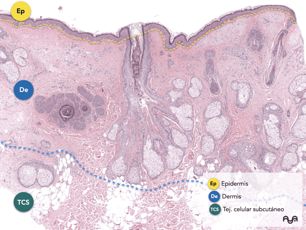
Key concepts
Normal dermis in one sentence:
It is the connective component of the skin that (1) mechanically supports the epidermis, (2) houses appendages and the neurovascular system, and (3) contains key cells for extracellular matrix (collagen/elastin/ground substance) generation and immune surveillance. Brown 2022
Papillary vs reticular dermis (the distinction you must master)
Papillary dermis
-
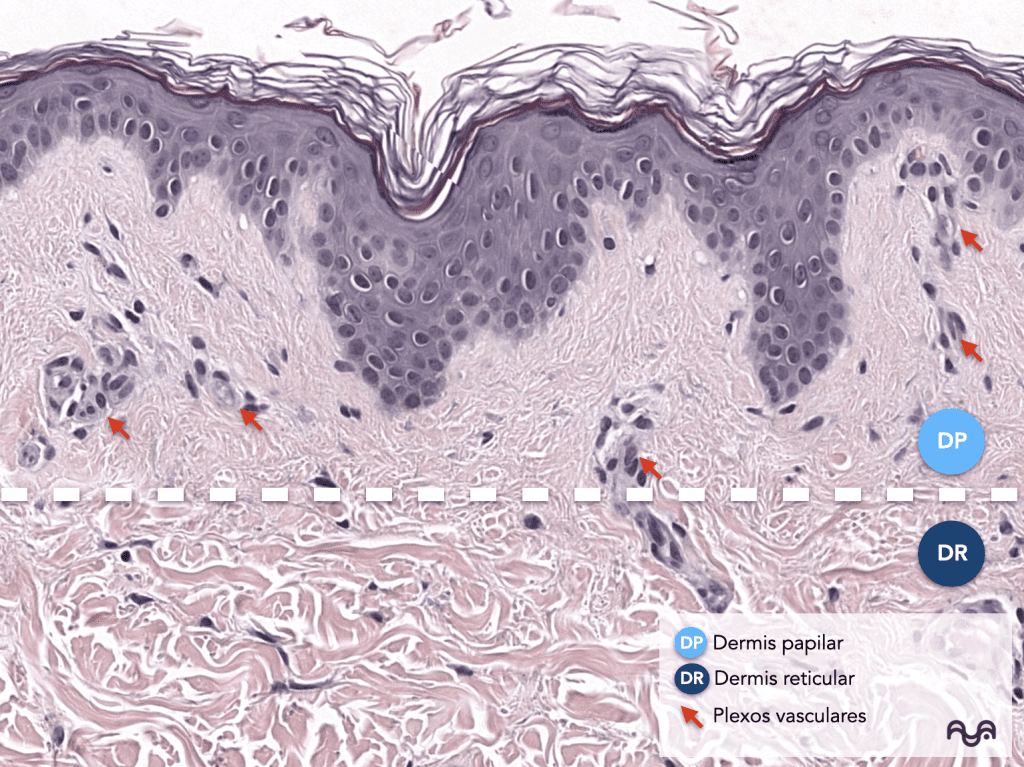
Looser , more cellular and more vascular connective tissue, adjacent to the dermoepidermal junction.
-
Wavy morphology due to dermal papillae (helps you orient yourself).
Brown 2022
Reticular dermis
-
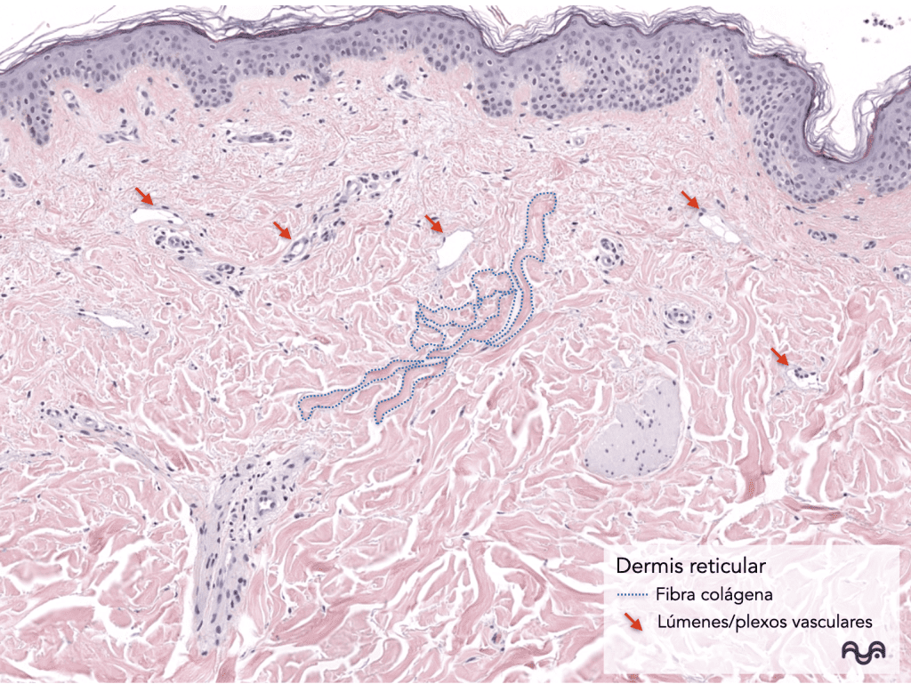
Dense irregular connective tissue: thicker , more eosinophilic collagen bundles with less apparent cellularity.
-
Deeper appendages and larger caliber vessels are more common here.
Brown 2022
Extracellular matrix: how it “translates” in H&E
-
Collagen: primarily responsible for eosinophilia and wavy bundles.
-
Elastin: it is not usually visible in standard H&E stains; however, it is important to consider it even if it does not «jump out» (it can be visualized with special stains for elastic fibers)
-
Ground substance (GAGs/proteoglycans): not seen as fiber; its presence is inferred from the background and by special techniques (e.g., Alcian blue for acidic mucins) Brown 2022
Microscopic evaluation (normal patterns + pearls)
“Layered” method (30–60 seconds)
-
Locate the epidermis to fix the «up».
-
Identify the most superficial (loosest) portion: it is the papillary dermis (papillae, fine capillaries).
-
Recognize the change to thick bundles: reticular dermis .
-
If fat appears, you are already in the hypodermis; the reticular dermis ends before the adipose lobules (if the cut includes that depth).
Agarwal 2023 ; Brown 2022
Practical tip: “How not to get lost with attachments”
-
If the follicle or a gland occupies the field, it relocates papillary near the junction; the appendages usually live in the reticular or in the reticular-hypodermis transition (depending on the site).
Agarwal 2023
Step-by-step approach (90 seconds)
Step 1 — Low magnification (2×–4×): map
-
Do you see the epidermis? Do you see the hypodermis? That defines the sampling depth.
-
Are there well-defined papillae or is the cut tangential?
-
What adnexal structures appear and on what plane?
Step 2 — Medium magnification (10×): define papillary and reticular
-
Papillary: lighter/looser connective tissue, smaller capillaries, more nuclei.
-
Reticular: collagen in thick bundles, fewer apparent nuclei, more frequent appendages.
Step 3 — High magnification (20×–40×): structural inventory
-
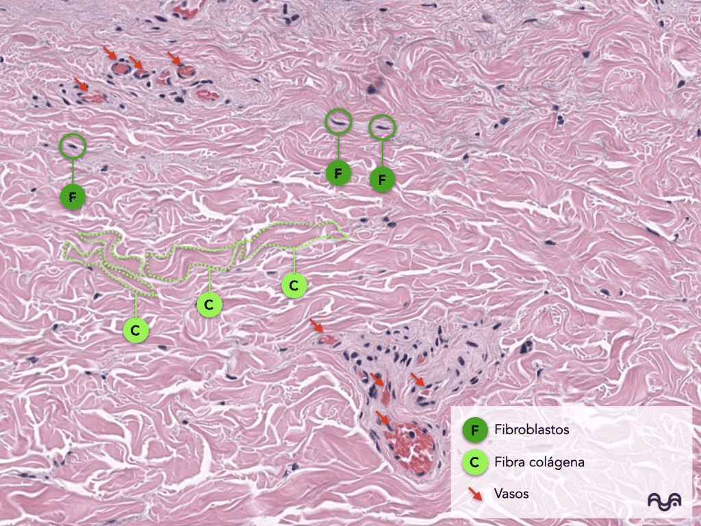
Vessels: lumen with erythrocytes, more defined wall (flat endothelium).
-
Nerves: wavy fascicles without lumen; subtle perineurium (sometimes).
-
Lymphatics: thin lumens, usually without erythrocytes; in H&E it may be subtle.
-
Fibroblasts: spindle-shaped nuclei, dispersed among collagen.
Brown 2022
Normality differentials (anatomical variation)
-
Anatomical site: the papillary gland may appear more or less prominent depending on the region (e.g., glabrous skin vs. hairy skin).
-
Dermal thickness: varies by region and by the presence of appendages.
-
Cutting orientation: can «flatten» papillae or exaggerate collagen bundles.
Agarwal 2023
Common Pitfalls (Technique and Reading)
-
Tangential sectioning: can cause papillary/reticular layers to become confused and collagen to appear «denser».
-
Retraction/shrinkage: artificial spaces around appendages or vessels.
-
Confusing nerve with smooth muscle (erector muscle): muscle is more eosinophilic and has elongated “cigar” nuclei; nerve has a paler/fibrillar and fasciculated appearance (rule of thumb; teacher confirmation with S100: link to be confirmed).
-
Mistaking a collapsed lymphatic for an artifact: look at continuity and relationship with vessels/stroma; in teaching, D2-40 may help (link to be confirmed).
-
Overinterpreting “cellularity”: papillary cells are relatively more cellular; not everything “more nuclei” is an abnormality.
Pearls, pitfalls, and common mistakes
-
If you cannot separate papillary/reticular, it is almost always due to the orientation of the cut : return to low magnification and look for papillae.
-
In H&E, elastin does not «compete» with collagen: if you want to study it, use elastic staining.
-
To locate receptors: think of “superficial = Meissner; deep = Pacini” as a rule of anatomical orientation Piccinin 2023
-
The appendages are part of the dermis: they don’t «break» your architecture; they just force you to reorient yourself.
Recommended readings
Essentials
-
Normal dermis: overview and papillary/reticular stratification — Brown 2022
-
Cutaneous anatomy/histology (context of dermis within skin) — Agarwal 2023
-
Meissner receptor (location and function) — Piccinin 2023
-
Epidermis (to anchor the upper boundary and layer orientation) — Yousef 2024
To delve deeper
-
Collagens in skin (emphasis on type I and III; includes normal skin bases) — Zhou 2024
-
Dermal elasticity and biogenesis of elastic fibers (mechanistic view) — Uitto 2012
-
Elastin in healthy skin and its structural relevance (review) — Baumann 2021
-
Extracellular matrix and dermal fibroblast (how they maintain ECM homeostasis) — Tracy 2016
-
Cutaneous microvasculature (papillary/reticular plexuses and capillary loops) — Deegan 2019
-
Morphology of vessels in skin and organization into plexuses (review) — Schuh 2017
-
Lymphatic vasculature: key structural concepts applicable to the dermis — Kerjaschki 2014
-
Elastic staining (Verhoeff–Van Gieson) to demonstrate elastic fibers (methodology and reading) — Percival 2016
Bibliography and sources
-
Brown TM, Krishnamurthy K. Histology, Dermis . StatPearls [Internet] . StatPearls Publishing; 2022. Brown 2022
-
Agarwal P, et al. Histology, Skin . StatPearls [Internet] . StatPearls Publishing; 2023. Agarwal 2023
-
Lopez-Ojeda W, et al. Anatomy, Skin (Integument) . StatPearls [Internet] . StatPearls Publishing; 2022. Lopez-Ojeda 2022
-
Yousef H, et al. Anatomy, Skin (Integument), Epidermis (for anchoring with the dermoepidermal junction). StatPearls [Internet] . 2024. Yousef 2024
-
Piccinin MA, et al. Histology, Meissner Corpuscle . StatPearls [Internet] . 2023. Piccinin 2023
-
Iheanacho F, et al. Physiology, Mechanoreceptors (includes Pacini in skin/subcutaneous). StatPearls [Internet] . 2023. Iheanacho 2023
-
Zhou CJ, et al. Mini review on collagens in normal skin and pathological scars . 2024. Zhou 2024
-
Ricard-Blum S. The Collagen Family . 2011. Ricard-Blum 2011
-
Uitto J, et al. The complexity of elastic fiber biogenesis in the skin . 2012. Uitto 2012
-
Baumann L, et al. Clinical Relevance of Elastin in the Structure and Function of Skin . 2021. Baumann 2021
-
Deegan AJ, et al. Microvascular Imaging of the Skin . 2019. Deegan 2019
-
Schuh S, et al. Imaging Blood Vessel Morphology in Skin . 2017. Schuh 2017
-
Geyer SH, et al. The dermal arteries of the human thumb pad . 2013. Geyer 2013
-
Kerjaschki D. The lymphatic vasculature revisited . 2014. Kerjaschki 2014
-
Hu Z, et al. Lymphatic vessel: origin, heterogeneity, biological functions… 2024. Hu 2024
-
Ter-Ovanesyan I, et al. An Update on the Role of Lymphatic Function in Skin… 2025. Ter-Ovanesyan 2025
Categories: Students and residents

Content reviewed by
Dr. Rodolfo Suárez
Médico Patólogo y Dermatólogo Master en Patología Cutánea Avanzada
- Published
- September 18, 2026
- Updated
- September 23, 2026
Medically reviewed by specialists
Our content is based on current scientific evidence and clinical protocols.