
Before you read: At Skinpaths, we distinguish between opinion and evidence. This content is based on indexed medical literature and recommendations from scientific societies. At the end, you will find a complete bibliography for verification and further study.
Learning objectives
-
Define the hypodermis (subcutis) and explain its relationship with the dermis and deep tissues (fascia). Lopez-Ojeda 2022 ; Fede 2025
-
Identify in H&E: adipocytes, lobules, fibrous septa and neurovascular bundles. Kim 2023 ; Esteve 2019
-
Recognize the existence of typical “deep” structures (e.g., Pacinian corpuscle) and locate them anatomically. Iheanacho 2023
-
Understanding the functions of the hypodermis (cushioning, insulation, energy reserve, and the endocrine role of adipose tissue). Kamrani 2023 ; Richard 2020
-
Avoid common errors in microscope interpretation (artifacts, orientation, and «false spaces»). Arregui 2020
Founding facts (what you absolutely must know)
-
The hypodermis is the deepest layer associated with skin, rich in adipose tissue and loose connective tissue , with larger blood vessels and nerves than in the dermis. Kim 2023
-
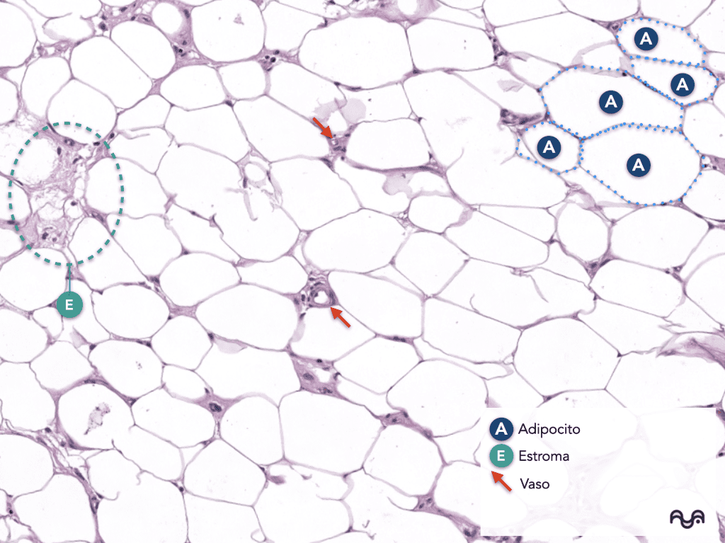
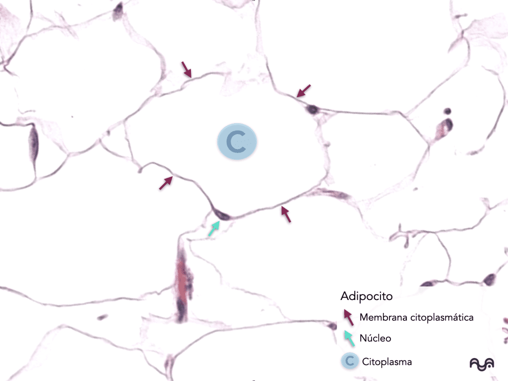
In H&E staining, the adipocyte is usually seen as a large optically empty space with a thin cytoplasmic rim : the fat is removed due to routine histological processing. Arregui 2020
-
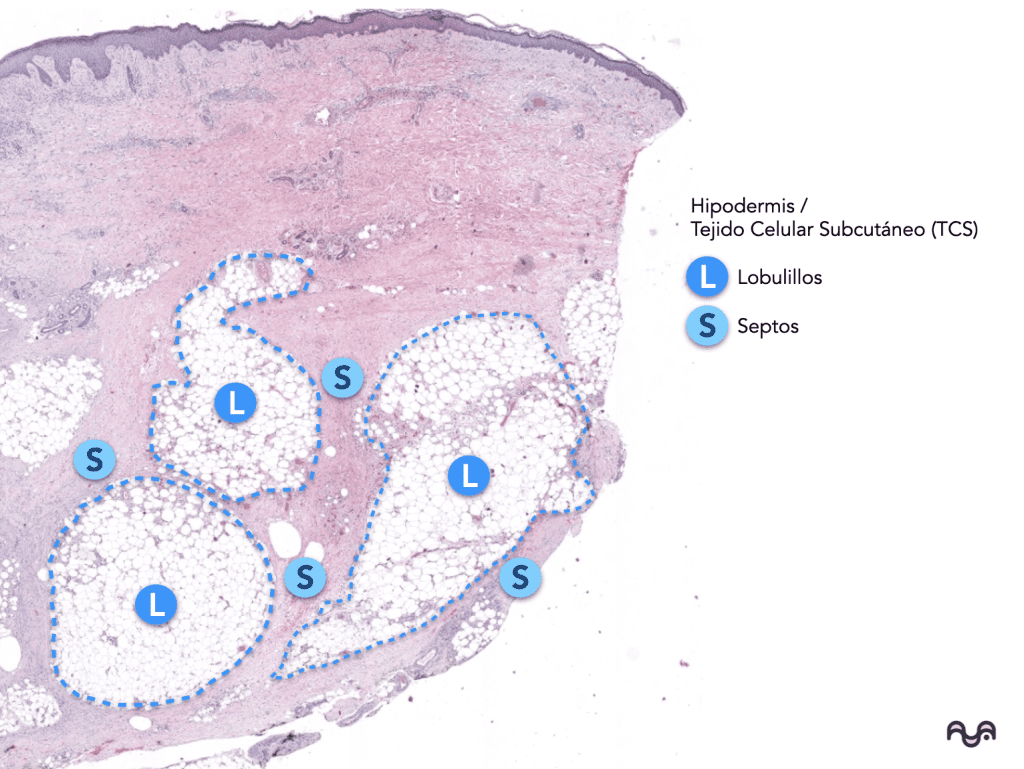
The typical architecture is lobular : fat lobules separated by fibrous septa (collagen) that «organize» the tissue. Estève 2019
-
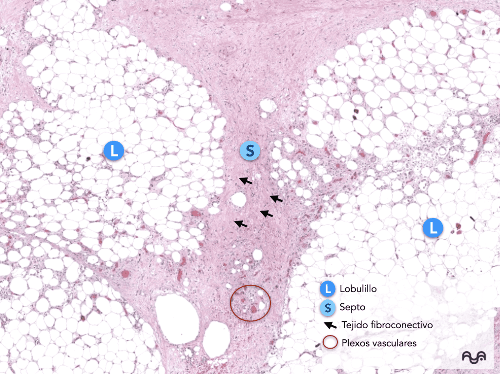
Septa often contain vessels , nerves , and, depending on the region, tracts of deep adnexa. Lopez-Ojeda 2022
-
The superficial fascia is described as a fibrous structure within the subcutaneous compartment; its nomenclature and boundaries may vary between texts. Fede 2025 ; Musil 2018
-
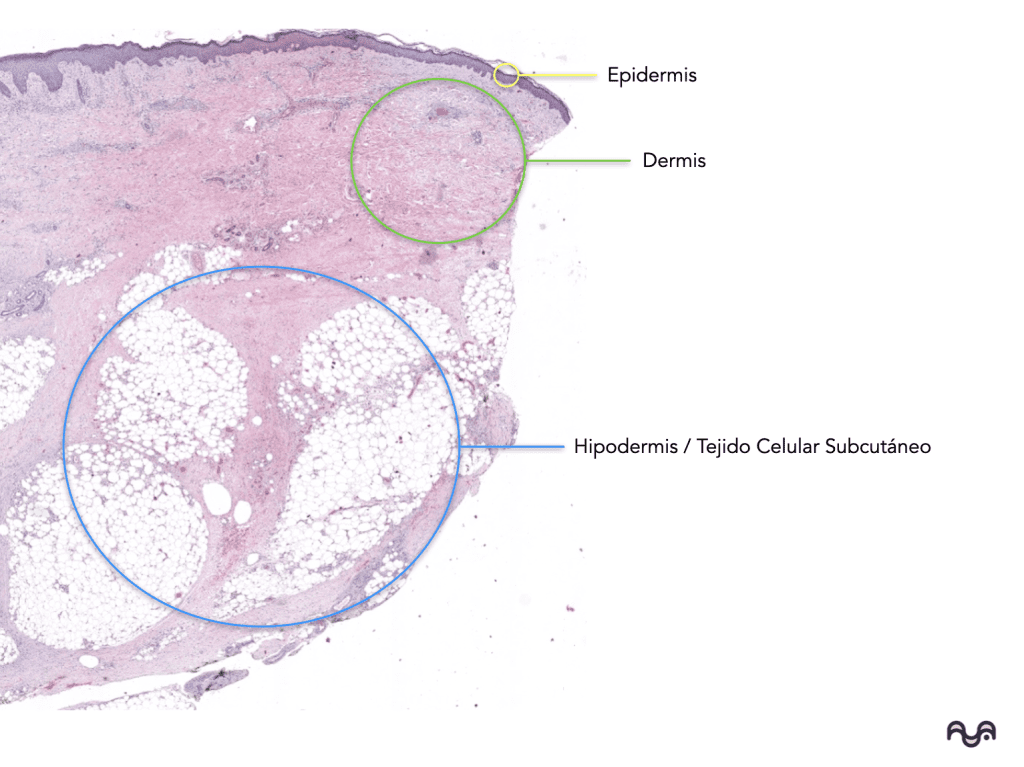
The transition from reticular dermis to hypodermis is usually evident: dense irregular collagen gives way to lobulated adipose «honeycomb» .
-
Pacinian corpuscles are large, encapsulated receptors, typically found in subcutaneous tissue (and deeper areas), with an » onion » pattern. Iheanacho 2023
-
Adipose tissue is not just for “filling”: it secretes mediators/hormones (adipokines) with systemic functions. Cinti 2022 ; Stern 2016
-
The hypodermis varies considerably by anatomical region and individual (thickness, lobular size, septal density). Fede 2025
-
In mammals, the panniculus carnosus (cutaneous muscle) is common; in humans, there are regional remnants (a useful concept as an “anatomical curiosity”). Naldaiz-Gastesi 2018 ; Musil 2018
-
If a biopsy does not reach the hypodermis (because it is superficial or because it was taken with an incorrect technique), you will not be able to «see» these clues: interpreting their «real absence» requires correlating with the depth of the sample .
Key concepts
Operational definition
The Hypodermis (Subcutaneous Cellular Tissue): This is the deep compartment beneath the dermis, composed of adipose and connective tissue , organized into lobules separated by fibrous septa . Kim 2023 ; Estève 2019
Main components
-
Adipocytes (white adipose tissue): specialized cells that store lipids; they are part of a dynamic organ with endocrine and paracrine functions. Richard 2020 ; Cinti 2022
-
Stroma/“stromal-vascular fraction”: the collection of non-adipocyte cells (endothelium, pericytes, fibroblasts, etc.) and extracellular matrix that supports the tissue. Cook 2009
-
Fibrous septa: connective tissue (collagen, variable elastic fibers) that compartmentalizes fat and serves as a «highway» for blood vessels and nerves. Estève 2019 ; Fede 2025
Origin (simplified)
-
Adipocytes are derived from mesenchymal progenitors through adipogenesis ; differences exist in development and biology depending on the type of fat deposit (subcutaneous vs. other). Berry 2013 ; Cook 2009
Microscopic presentation (how to see and “read” it)
1) At low magnification (2×–4×): “layer map”
-
Identify the reticular dermis: a relatively homogeneous, dense pink band (collagen).
-
Look for the change in texture: a clear lobed pattern appears (“honeycomb”): that is the hypodermis .
-
Identify septa: eosinophilic lines/bands that separate groups of “lobes”.
Pearl: If the cut is tangential or the biopsy is superficial, you could be in a scenario where you would only identify deep dermis (without hypodermis).
2) At intermediate magnification (10×): “lobule vs septum”
-
Adipose lobule: a cluster of adipocytes with little connective tissue between them.
-
Septum: denser connective tissue, containing vessels (lumen + wall lined with endothelial cells) and sometimes nerves (wavy bundles with a subtle perineurium). Lopez-Ojeda 2022
3) At high magnification (20×–40×): “adipocyte signature”
-
Typical white adipocyte: large vacuole (lipid extracted by histological processing) + thin peripheral cytoplasm ; the nucleus may be difficult to see in suboptimal sections.
-
What appears to be “space” is actually the imprint of the lipid lost by solvents during histological processing. Arregui 2020
Structures that may appear nearby
-
Deep glandular structures (secretory portion of eccrine glands) are usually in the deep dermis and may «touch» the boundary with the hypodermis.
-
Hair follicles : In some regions, deep structures of the follicle may extend into nearby planes.
Pitfalls (common mistakes)
-
Confusing processing “gaps” with cystic structures or pathological spaces: first think of extracted fat . Arregui 2020
-
Not recognizing the septum : if you only look at high magnification, you may miss the lobular architecture.
-
Overinterpreting “absence of hypodermis” when the biopsy is superficial.
Recommended readings
Essentials
-
General anatomy of the skin (includes subcutis/hypodermis) — Lopez-Ojeda 2022
-
Physiology of the integument (functions of the subcutaneous tissue) — Kim 2023
-
Connective tissue and adipose tissue as specialized connective tissue — Kamrani 2023
-
Adipose tissue: biology and function (NCBI chapters) — Richard 2020
-
Deep cutaneous receptors (Pacinian) — Iheanacho 2023
To delve deeper
-
Lobular architecture of subcutaneous adipose tissue and septa — Estève 2019
-
Origin/development and heterogeneity of adipose deposits — Berry 2013
-
The human superficial fascia: components, function and anatomical discussion — Fede 2025
-
Superficial fascia system and its relationship with subcutaneous morphology — Si 2022
-
Adipose tissue as an endocrine organ (integrative view) — C inti 2022
-
Leptin and signaling from adipose tissue (example of adipokine) — Martínez-Sánchez 2020
-
Histological techniques to demonstrate fat (OsO₄ vs Oil Red O, etc.) — Arregui 2020
-
Panniculus carnosus: anatomical/evolutionary review (context for “subcutaneous + musculocutaneous”) — Naldaiz-Gastesi 2018
Bibliography and sources
-
Lopez-Ojeda W, Hurley S. Anatomy, Skin (Integument). StatPearls Publishing; 2022. Lopez-Ojeda 2022
-
Kim JY, et al. Physiology, Integument. StatPearls Publishing; 2023. Kim 2023
-
Kamrani P, et al. Anatomy, Connective Tissue. StatPearls Publishing; 2023. Kamrani 2023
-
Richard AJ, et al. Adipose Tissue: Physiology to Metabolic Dysfunction. In: Endotext/NCBI Bookshelf ; 2020. Richard 2020
-
Cook A, Cowan C. Adipose. StemBook/NCBI Bookshelf; 2009. Cook 2009
-
Berry DC, Stenesen D, Zeve D, Graff JM. The developmental origins of adipose tissue. Development. 2013.Berry 2013
-
Estève D, et al. Lobular architecture of human adipose tissue defines the niche and fate of progenitor cells. Nat Commun. 2019. Esteve 2019
-
Fede C, et al. The Human Superficial Fascia: A Narrative Review. Int J Mol Sci. 2025. Fede 2025
-
If L, et al. The Superficial Fascia System: Anatomical Guideline for Subcutaneous Fat Morphology. Aesthet Surg J Open Forum. 2022. Yes 2022
-
Cinti F. The Endocrine Adipose Organ: A System Playing a Central Role in Metabolism and Inflammation. Front Endocrinol. 2022. Cinti 2022
-
Stern JH, Rutkowski JM, Scherer PE. Adiponectin, Leptin, and Fatty Acids in the Maintenance of Metabolic Homeostasis. Trends Endocrinol Metab. 2016. Stern 2016
-
Martínez-Sánchez N, et al. There and Back Again: Leptin Actions in White Adipose Tissue. Front Endocrinol. 2020. Martínez-Sánchez 2020
-
Iheanacho F, et al. Physiology, Mechanoreceptors. StatPearls Publishing; 2023. Iheanacho 2023
-
Arregui M, et al. Comparison of Three Histological Techniques for Fat Demonstration: Osmium Tetroxide, Chromic Acid, and Oil Red O. Sci Rep. 2020. Arregui 2020
-
Du J, et al. An optimized method for Oil Red O staining with the removal of background interference. Cell Biol Int. 2023. Du 2023
-
Melo RCN, et al. Imaging Lipid Bodies Within Leukocytes… (osmium staining section). Methods Mol Biol. 2011. Melo 2011
-
Naldaiz-Gastesi N, et al. The panniculus carnosus muscle: an evolutionary enigma… Dev Biol. 2018. Naldaiz-Gastesi 2018
-
Musil V, et al. A plea for an extension of the anatomical nomenclature (fascia superficialis and relationship with cutaneous muscle). J Anat. 2018. Musil 2018
Categories: Students and residents

Content reviewed by
Dr. Rodolfo Suárez
Médico Patólogo y Dermatólogo Master en Patología Cutánea Avanzada
- Published
- September 18, 2026
- Updated
- September 23, 2026
Medically reviewed by specialists
Our content is based on current scientific evidence and clinical protocols.