
Before you read: At Skinpaths, we distinguish between opinion and evidence. This content is based on indexed medical literature and recommendations from scientific societies. At the end, you will find a complete bibliography for verification and further study.
Learning objectives
-
Recognize the layered architecture in H&E: epidermis → dermoepidermal junction → dermis → hypodermis.
-
Identify the epidermal layers and their morphological “clues”.
-
Understanding what the basement membrane zone (dermoepidermal junction) is and why it matters.
-
Differentiate papillary vs reticular dermis using reproducible criteria.
-
Locate key cells (keratinocyte, melanocyte, Langerhans cell, fibroblast, adipocyte) and their function.
-
Apply a systematic reading to avoid missing normal variations (anatomical site, thick/thin skin, appendages).
Founding facts (what you absolutely must know)
-
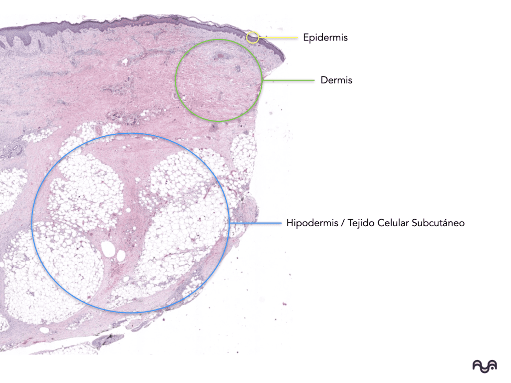
The skin is organized into the epidermis (keratinized stratified squamous epithelium), dermis (connective tissue), and hypodermis (lobular adipose tissue with septa). Agarwal 2023
-
The epidermis is avascular ; it is nourished by diffusion from the dermis through the dermoepidermal junction. Yousef 2024
-
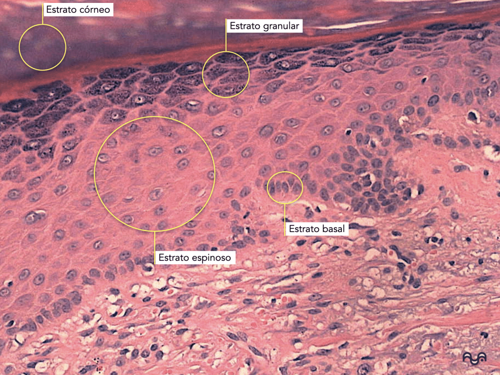
Epidermal layers (superficial → deep): corneous, granular, spinous, basal.
The stratum lucidum is only present in thick skin (palms/soles). Kim 2023
-
In H&E, the granular layer stands out for basophilic keratohyalin granules ; the spinous layer for “ intercellular bridges ” (desmosomes).
-
The keratinocyte dominates the epidermis and completes cornification to form the stratum corneum (physical barrier). Gutowska-Owsiak 2020
-
The stratum corneum barrier is understood as “ bricks and mortar ”: corneocytes (bricks) + lamellar lipids (mortar). Del Rosso 2016
-
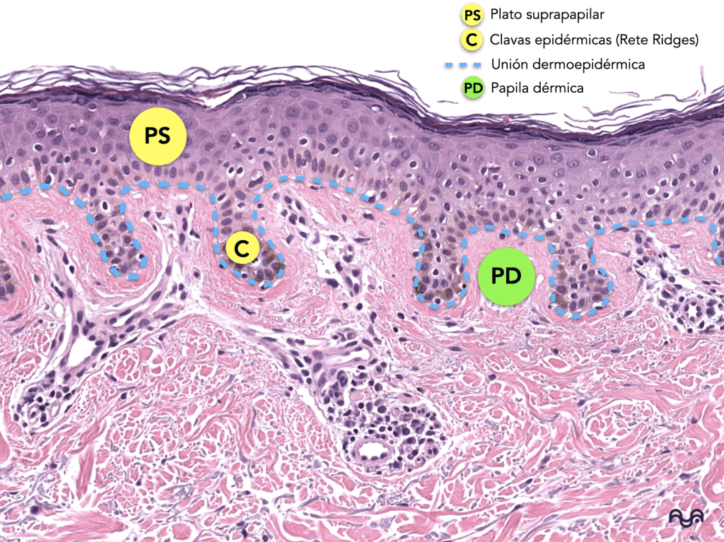
The dermoepidermal junction (basement membrane zone) is an adhesion superstructure ; its integrity depends on laminin-332, collagen IV, perlecan , and interactions with integrins/hemidesmosomes. Kiritsi 2013 , Behrens 2012
-
Anchoring filaments and anchoring fibrils (collagen VII) connect the epidermis to the papillary dermis: a “critical zone” for subepidermal blisters. Goletz 2017 , Villone 2008
-
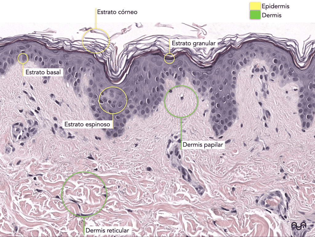
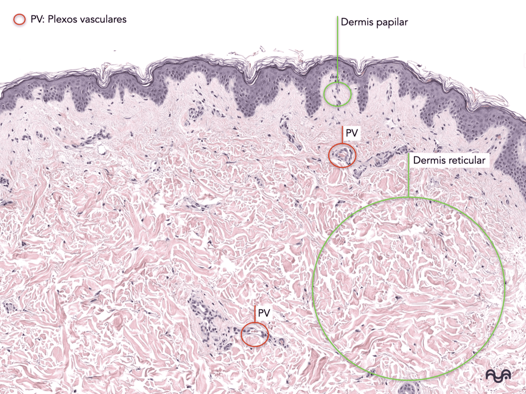
The dermis is divided into papillary (loose connective tissue, more vascular, with papillae) and reticular (dense irregular connective tissue, with thick bundles of collagen). Brown 2022
-
In the papillary dermis, Meissner’s corpuscles (fine touch) can be found in glabrous skin; in the deep dermis/hypodermis, Pacinian corpuscles (vibration/pressure). Piccinin 2023 Brown 2022
-
Melanocytes (origin: neural crest ) reside primarily in the basal layer and transfer melanosomes to keratinocytes. Huang 2024
-
Langerhans cells (myeloid lineage) reside mainly in the stratum spinosum and act as immunological sentinels at the epidermal level. Igyártó 2013
-
Merkel cells (form complexes with afferent fibers for discriminative touch; they are usually located in the basal epidermis/“touch domes”. Woo 2015
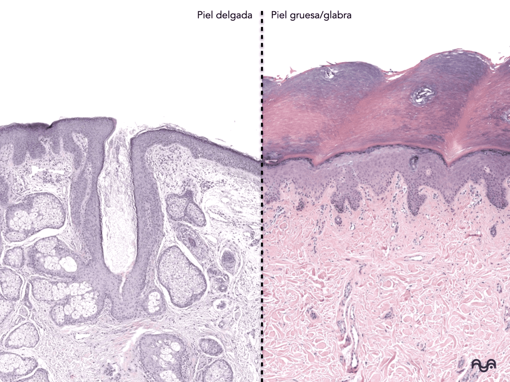
Thick skin : stratum corneum very developed, lucid present , without hair follicles or sebaceous glands , with many eccrine glands . Lopez-Ojeda 2022
Thin skin : thinner stratum corneum, with follicles and pilosebaceous units that vary depending on the site. Agarwal 2023
Key concepts
1) General plan (what you are seeing on the portal)
-
Epidermis: stratified squamous keratinized epithelium, with continuous renewal from the basal layer towards the surface (terminal differentiation/cornification). Gutowska-Owsiak 2020
-
Dermis: connective tissue of mesenchymal origin (mesoderm), providing mechanical support, immunity, and repair; extracellular matrix rich in collagen/elastin and ground substance. Brown 2022
-
Hypodermis (subcutaneous cellular tissue): adipocyte lobules with fibrous septa; cushioning, energy reserve, thermal insulation, and endocrine functions of adipose tissue. Yousef 2024 Kershaw 2004
2) Normal variation (why it “doesn’t look the same” in all cuts)
-
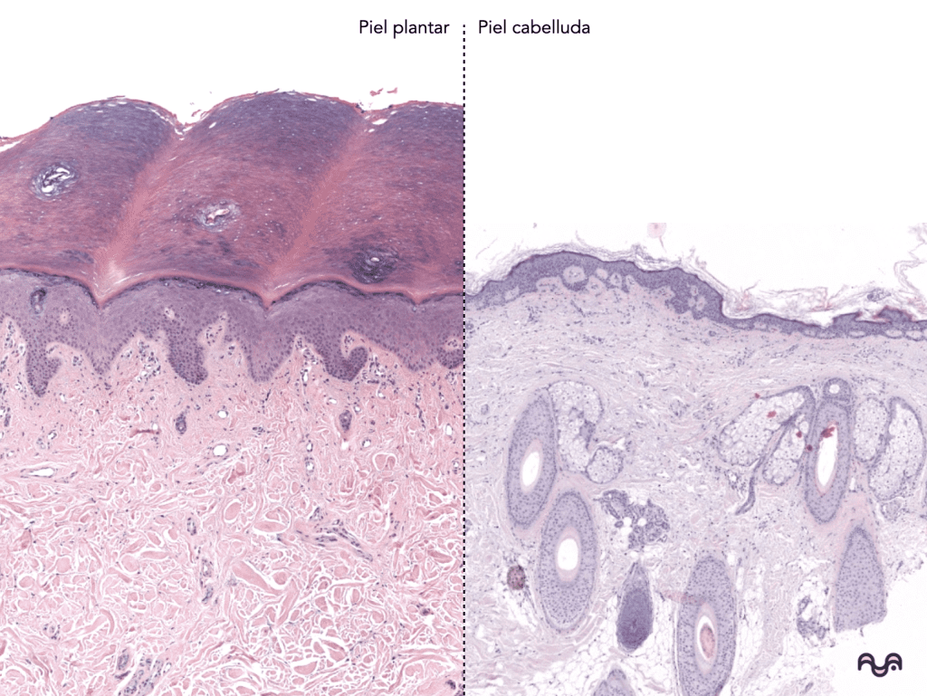
Anatomical site: palms/soles (glabrous) vs scalp vs trunk.
-
Cutting orientation: perpendicular vs tangential (artifacts).
-
Age/photoexposure: modifies epidermal thickness, elastosis, and adnexal density (general concept; for future clinical correlation).
-
Functional state of the follicle: anagen/catagen/telogen (morphology changes).
Histological presentation of normal skin (according to its anatomical zone)
-
Glabrous skin (palm/sole): very thick epidermis, prominent epidermal ridges, massive stratum corneum, abundant eccrine glands; absence of pilosebaceous unit. Lopez-Ojeda 2022
-
Hairy skin (most of the body): hair follicles in different planes, associated sebaceous glands, arrector pili muscle, variable eccrine glands. Agarwal 2023
-
Papillary dermis: “lighter” and looser near the junction; capillaries in loops within papillae.
-
Reticular dermis: thicker collagen bundles, more eosinophilic, with deeper appendages.
-
Perla: When you get «lost,» return to two visual anchors : (1) stratum corneum above; (2) fat (if present) below. Everything else is organized between these references.
Systematic skin reading exercise
“Top-down” method (60–90 second method)
Step 1 — Low magnification (2×–4×): general map
-
Identify the surface : eosinophilic stratum corneum (sometimes laminated).
-
Decide if it’s thick skin vs thin skin :
-
-
thick: very wide stratum corneum + (possible) stratum lucidum + without follicles;
-
Thin: thin stratum corneum + follicles with or without hair in the lumen. Kim 2023
-
-
Locate the appendages : follicles, glands, ducts, and arrector pili muscle. Agarwal 2023
-
Look for the hypodermis (if present): fat lobules with septa.
Step 2 — Medium magnification (10×): epidermis by layers
-
Basal layer: a row of cuboidal/low columnar cells with basophilic nuclei and scant cytoplasm. This layer is where melanocytes reside (not always obvious in H&E staining).
-
Spinous: polygonal keratinocytes; “spines” due to desmosomes (retraction artifact that highlights intercellular bridges).
-
Granular: basophilic keratohyalin granules; marks the “threshold” towards cornification.
-
Corneum: anucleate corneocytes; flattened on their horizontal axis and of variable compaction (normal orthokeratosis). Gutowska-Owsiak 2020
Step 3 — The dermoepidermal junction (the critical line)
-
In H&E it looks like a wavy outline : epidermal ridges (rete ridges) that go down and dermal papillae that go up.
-
Conceptually, this is the basement membrane zone : laminin-332 and collagen IV form networks; perlecan contributes to organization; hemidesmosomes anchor the basal keratinocyte. Kiritsi 2013, Behrens 2012, Walko 2014
Step 4 — Dermis: papillary vs reticular
-
Papillary: looser, more cellular and vascular connective tissue; papillae; possibly Meissner’s papillae in glabrous skin. Brown 2022
-
Reticular: thick bundles of collagen (dense irregular connective tissue), fewer apparent cells, deeper appendages.
Step 5 — Hypodermis
-
Unilocular adipocytes: “empty” cytoplasm (extracted lipid) with a peripheral nucleus; septa with vessels/nerves. Yousef 2024
Useful differences (when you’re unsure if «it’s leather»)
-
Non-keratinized squamous mucosa : absence of a true stratum corneum.
-
Skin vs scalp : high follicular density and prominent pilosebaceous units.
-
Glabrous vs thin skin : absence/presence of follicles and sebaceous glands.
Pitfalls (typical mistakes)
-
Tangential section : may simulate acanthosis/atrophy; confirm with the global pattern at low magnification.
-
Retraction : spaces at the junction or between keratinocytes may look like «spongiosis» if you don’t assess the context.
-
Follicle planes : a follicle in cross-section may look like a “cyst”; look for the rest of the unit and pattern repetition.
-
Confusing the stratum lucidum with the «compact stratum corneum»: the stratum lucidum is a pale band between the granular and corneum layers, typical of thick skin. Kim 2023
-
Calling any tubular structure a «gland» distinguishes the eccrine duct (darker, bi-layered epithelium) from the secretory portion (lighter/more varied and myoepithelial cells). Yousef 2023
-
Thinking that “no fat = no skin”: there are superficial biopsies without hypodermis; look for appendages and reticular dermis.
Recommended readings
Essentials
To delve deeper
Bibliography and sources
-
Yousef H, Alhajj M, Fakoya AO, Sharma S. Anatomy, Skin (Integument), Epidermis . In: StatPearls [Internet] . Treasure Island (FL): StatPearls Publishing; 2025 Jan–. Updated: Jun 8, 2024. Available at: https://www.ncbi.nlm.nih.gov/books/NBK470464/
-
Yousef H, Miao JH, Alhajj M, Badri T. Histology, Skin Appendages . In: StatPearls [Internet] . Treasure Island (FL): StatPearls Publishing; 2025 Jan–. Updated: Apr 24, 2023. Available at: https://www.ncbi.nlm.nih.gov/books/NBK482237 /
-
Brown TM, Krishnamurthy K. Histology, Hair and Follicle . In: StatPearls [Internet] . Treasure Island (FL): StatPearls Publishing; 2025 Jan–. Updated: May 1, 2023. Available at: https://www.ncbi.nlm.nih.gov/books/NBK532929 /
-
Piccinin MA, Miao JH, Schwartz J. Histology, Meissner Corpuscle . In: StatPearls [Internet] . Treasure Island (FL): StatPearls Publishing; 2025 Jan–. Updated: Mar 6, 2023. Available at: https://www.ncbi.nlm.nih.gov/books/NBK518980/
-
Patel BC, Treister AD, McCausland C, Lio PA, Jozsa F. Anatomy, Skin, Sudoriferous Gland . In: StatPearls [Internet] . Treasure Island (FL): StatPearls Publishing; 2025 Jan–. Updated: Apr 24, 2023. Available at: https://www.ncbi.nlm.nih.gov/books/NBK513244/
-
Freeman SC, Malik A, Basit H. Physiology, Exocrine Gland . In: StatPearls [Internet] . Treasure Island (FL): StatPearls Publishing; 2025 Jan–. Updated: Sep 26, 2022. Available at: https://www.ncbi.nlm.nih.gov/books/NBK542322/
-
Gutowska-Owsiak D, et al. Addressing Differentiation in Live Human Keratinocytes by Assessment of Membrane Packing Order . Front Cell Dev Biol . 2020;8:573230. doi:10.3389/fcell.2020.573230. Available at: https://pmc.ncbi.nlm.nih.gov/articles/PMC7609878/
-
Bikle DD, Xie Z, Tu CL. Calcium regulation of keratinocyte differentiation . Expert Rev Endocrinol Metab . 2012;7(4):461–472. doi:10.1586/eem.12.34. Available at: https://pmc.ncbi.nlm.nih.gov/articles/PMC3491811/
-
Del Rosso JQ, et al. Understanding the Epidermal Barrier in Healthy and Compromised Skin: Clinically Relevant Information for the Dermatology Practitioner. Proceedings of an Expert Panel Roundtable Meeting . J Clin Aesthet Dermatol . 2016;9(4 Suppl 1):S2–S8. Available at: https://pmc.ncbi.nlm.nih.gov/articles/PMC5608132/
-
Kiritsi D, Has C, Bruckner-Tuderman L. Laminin 332 in junctional epidermolysis bullosa . Cell Adh Migr . 2013;7(1):135–141. doi:10.4161/cam.22418. Available at: https://pmc.ncbi.nlm.nih.gov/articles/PMC3544777/
-
Behrens DT, et al. The Epidermal Basement Membrane Is a Composite of Separate Laminin- or Collagen IV-containing Networks Connected by Aggregated Perlecan, but Not by Nidogens . J Biol Chem . 2012;287(22):18700–18709. doi:10.1074/jbc.M111.336073. Available at: https://pmc.ncbi.nlm.nih.gov/articles/PMC3365761/
-
Walko G, Castañón MJ, Wiche G. Molecular architecture and function of the hemidesmosome . Cell Tissue Res . 2014;360(2):363–378. doi:10.1007/s00441-014-2061-z. Available at: https://pmc.ncbi.nlm.nih.gov/articles/PMC4544487/
-
Goletz S, Zillikens D, Schmidt E. Structural proteins of the dermal-epidermal junction targeted by autoantibodies in pemphigoid diseases . Exp Dermatol . 2017;26(12):1154–1162. doi:10.1111/exd.13446. Available at: https://pubmed.ncbi.nlm.nih.gov/28887824/
-
Villone D, et al. Supramolecular interactions in the dermo-epidermal junction zone: anchoring fibril-collagen VII tightly binds to banded collagen fibrils . J Biol Chem . 2008;283(36):24506–24513. doi:10.1074/jbc.M802415200. Available at: https://pubmed.ncbi.nlm.nih.gov/18599485/
-
Niemann C, Horsley V. Development and homeostasis of the sebaceous gland . Semin Cell Dev Biol . 2012;23(8):928–936. doi:10.1016/j.semcdb.2012.08.010. Available at: https://pmc.ncbi.nlm.nih.gov/articles/PMC5595243/
-
Cui CY, Schlessinger D. Eccrine sweat gland development and sweat secretion . Exp Dermatol . 2015;24(9):644–650. doi:10.1111/exd.12773. Available at: https://pmc.ncbi.nlm.nih.gov/articles/PMC5508982/
-
Igyártó BZ, Kaplan DH. Antigen presentation by Langerhans cells . Curr Opinion Immunol . 2013;25(1):115–119. doi:10.1016/j.coi.2012.11.007. Available at: https://pubmed.ncbi.nlm.nih.gov/23246038/
-
Woo SH, Lumpkin EA, Patapoutian A. Merkel cells and neurons keep in touch . Trends Cell Biol . 2015;25(2):74–81. doi:10.1016/j.tcb.2014.10.003. Available at: https://pubmed.ncbi.nlm.nih.gov/25480024/
-
Bento-Lopes L, et al. Melanin’s Journey from Melanocytes to Keratinocytes: Uncovering the Molecular Mechanisms of Melanin Transfer and Processing . Int J Mol Sci . 2023;24(14):11289. doi:10.3390/ijms241411289. Available at: https://pmc.ncbi.nlm.nih.gov/articles/PMC10379423/
-
Vandamme N, et al. From neural crest cells to melanocytes: cellular plasticity during development and beyond . Cell Mol Life Sci . 2019;76(10):1919–1934. doi:10.1007/s00018-019-03049-w. Available at: https://pmc.ncbi.nlm.nih.gov/articles/PMC11105195/
-
Huang L, Zuo Y, Li S, Li C. Melanocyte stem cells in the skin: Origin, biological characteristics, homeostatic maintenance and therapeutic potential . Clin Transl Med . 2024;14(5):e1720. doi:10.1002/ctm2.1720. Available at: https://pubmed.ncbi.nlm.nih.gov/38778457/
-
Kershaw EE, Flier JS. Adipose tissue as an endocrine organ . J Clin Endocrinol Metab . 2004;89(6):2548–2556. doi:10.1210/jc.2004-0395. Available at: https://pubmed.ncbi.nlm.nih.gov/15181022/
Tags
Categories: Students and residents

Content reviewed by
Dr. Rodolfo Suárez
Médico Patólogo y Dermatólogo Master en Patología Cutánea Avanzada
- Published
- September 18, 2026
- Updated
- September 23, 2026
Medically reviewed by specialists
Our content is based on current scientific evidence and clinical protocols.