

A practical guide for dermatologists on when and how to perform a biopsy in patients with alopecia, focusing on the aspects that most influence diagnostic performance.
Before reading: This article is intended exclusively for healthcare professionals (physicians, residents, and other medically trained professionals). The content is for educational and academic updating purposes and does not replace individual clinical assessment, professional judgment, or specialized medical consultation . Diagnostic and therapeutic decisions should always be made within the context of each individual patient and under the responsibility of the treating physician.
The information presented is based on concepts widely described in current scientific literature, clinical guidelines, and articles published in specialized medical journals . However, medicine is a constantly evolving field, so the content presented here should not be interpreted as absolute truth or a universal recommendation applicable to all cases . The reader should consider this information as a general guideline and is always advised to review the original sources cited , as well as the most recent available evidence, before making specific clinical decisions.
1. When is a biopsy worthwhile?
Consider implementing it when the result may change therapeutic behavior :
-
Actual diagnostic uncertainty after history, examination, and trichoscopy (especially if it is among several non-scarring alopecias). [show_150ms_ease-in] » data-testid=»webpage-citation-pill»> PubMed
-
Suspected primary scarring alopecia (LPP, FFA, DLE, folliculitis decalvans, etc.): biopsy is practically mandatory. [show_150ms_ease-in] » data-testid=»webpage-citation-pill»> onlinelibrary.wiley.com
-
Chronic diffuse alopecia in women , with uncertainty between chronic telogen effluvium, androgenetic alopecia, mixed causes, or systemic causes. [show_150ms_ease-in] » data-testid=»webpage-citation-pill»> British Hair and Nail Society
-
Atypical/diffuse/incognita alopecia areata , or possible coexistence with effluvium/AGA. [show_150ms_ease-in] » data-testid=»webpage-citation-pill»> pmc.ncbi.nlm.nih.gov
-
Failure of successive treatments without a clear diagnosis, before escalating to immunosuppressants, retinoids, JAK, etc. [show_150ms_ease-in] » data-testid=»webpage-citation-pill»> PubMed
2. Three key decisions before taking the biopsy
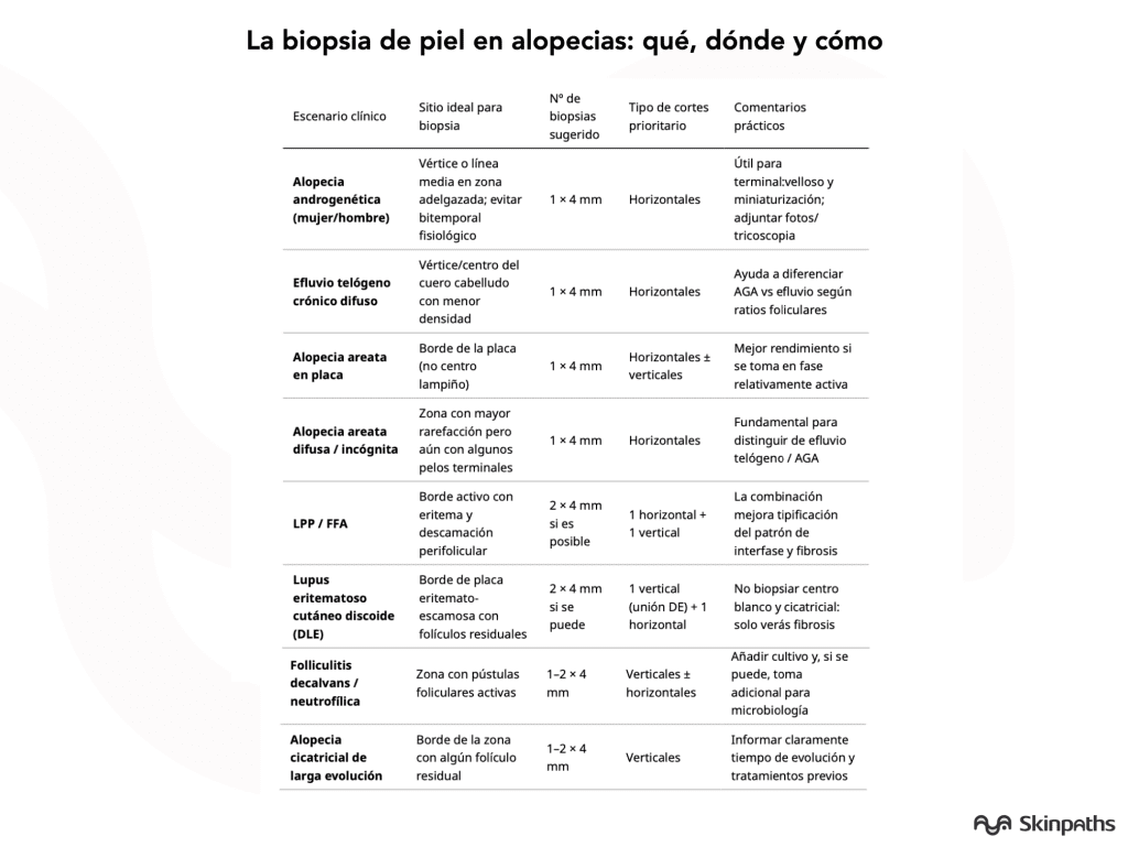
2.1. Choosing the right site
General rule of thumb: biopsy active area , not totally scarred tissue.
-
Non-scarring alopecia (AGA, effluvium, AA):
-
Prefer vertex or representative mid-parietal region .
-
Always avoid the bitemporal region (physiological miniaturization can be confusing). [show_150ms_ease-in] » data-testid=»webpage-citation-pill»> British Hair and Nail Society
-
-
Androgenetic alopecia (woman or man):
-
Area of greatest thinning in the midline (not extreme frontal border). [show_150ms_ease-in] » data-testid=»webpage-citation-pill»> anddermatol.org
-
-
Chronic telogen effluvium:
-
Vertex or central zone where a diffuse decrease in density is perceived.
-
Avoid areas with previous scarring, radiation therapy, or traction. [show_150ms_ease-in] » data-testid=»webpage-citation-pill»> PubMed
-
-
Alopecia areata in plaque:
-
The edge of the plaque, where miniaturized/damaged follicles still remain, not the completely hairless center. [show_150ms_ease-in] » data-testid=»webpage-citation-pill»> pmc.ncbi.nlm.nih.gov
-
-
Primary cicatricial alopecias (LPP/FFA, DLE, FD, etc.):
-
Active inflammatory border (perifollicular erythema, scaling, pustules, pain or itching).
-
Avoid the smooth, white center, which will only show fibrosis. [show_150ms_ease-in] » data-testid=»webpage-citation-pill»> PubMed
-
2.2. How many biopsies?
-
A 4 mm biopsy is usually sufficient in non-scarring alopecias if the pathologist is experienced and makes horizontal sections. [show_150ms_ease-in] » data-testid=»webpage-citation-pill»> ishrs-htforum.org
-
In primary scarring alopecias , the ideal (if the scalp allows it) is:
-
Two 4 mm punches : one for horizontal cuts and one for vertical cuts. [show_150ms_ease-in] » data-testid=»webpage-citation-pill»> Plastic Surgery Key
-
2.3. Orientation and depth
-
Size: punch 4 mm (current standard). [show_150ms_ease-in] » data-testid=»webpage-citation-pill»> British Hair and Nail Society
-
Depth: down to the subcutaneous tissue ; if it remains in the mid-dermis, the bulb is not visible. [show_150ms_ease-in] » data-testid=»webpage-citation-pill»> pathology.alfred.org.au
-
Angle: Follow the hair shaft to avoid massive follicle transection. [show_150ms_ease-in] » data-testid=»webpage-citation-pill»> journals.lww.com
-
Communication with the pathologist: always indicate:
-
Exact location.
-
Pattern type (diffuse, plaque-like, frontal, scarring/non-).
-
Time of evolution, previous treatments and trichoscopy.
-
Differential diagnoses being considered. [show_150ms_ease-in] » data-testid=»webpage-citation-pill»> PubMed
-
3. Horizontal vs. vertical cuts: what to ask for and when
3.1. Horizontal (cross-sections)
-
They allow for counting follicles and studying:
-
Terminal:villous ratio (miniaturization).
-
Anagen:telogen ratio.
-
Follicular distribution per unit. [show_150ms_ease-in] » data-testid=»webpage-citation-pill»> pmc.ncbi.nlm.nih.gov
-
-
They are especially useful in:
-
AGA (miniaturization pattern). [show_150ms_ease-in] » data-testid=»webpage-citation-pill»> Nature
-
Chronic telogen effluvium . [show_150ms_ease-in] » data-testid=»webpage-citation-pill»> PubMed
-
Alopecia areata , especially in the intermediate phases between acute and chronic outbreaks. [show_150ms_ease-in] » data-testid=»webpage-citation-pill»> onlinelibrary.wiley.com
-
3.2. Vertical (longitudinal) cuts
-
Ideal for evaluation:
-
Dermoepidermal junction , interface changes.
-
Pattern of fibrosis and follicular destruction.
-
Depth of perifollicular inflammation. [show_150ms_ease-in] » data-testid=»webpage-citation-pill»> PubMed
-
-
Very valuable in:
-
LPP/FFA , DLE , folliculitis decalvans and other scarring. [show_150ms_ease-in] » data-testid=»webpage-citation-pill»> PubMed
-
3.3. What does the evidence/committees say?
-
Comparative studies show that horizontal and vertical alignments complement each other ; the combination is superior to either one separately. [show_150ms_ease-in] » data-testid=»webpage-citation-pill»> Journal of Laboratory Physicians
-
If only one biopsy can be sent, many guidelines suggest:
-
Prioritize horizontal haircuts in non-scarring alopecia. [show_150ms_ease-in] » data-testid=»webpage-citation-pill»> PubMed
-
Consider vertical cuts if the main suspicion is PPL/FFA or other interface scarring . [show_150ms_ease-in] » data-testid=»webpage-citation-pill»> Journal of Laboratory Physicians
-
4. Trichoscopy + biopsy: how they complement each other
-
Trichoscopy is now a standard tool for differentiating patterns and choosing the best biopsy site. [show_150ms_ease-in] » data-testid=»webpage-citation-pill»> journals.viamedica.pl
-
Practical examples:
-
LPP/FFA: perifollicular scale + erythema → active border for biopsy.
-
FD: follicular pustules and hemorrhagic crusts → area with intense inflammation.
-
AGA vs effluvium: heterogeneous miniaturization + hairy hairs → prioritize area with clearer findings.
-
-
In some non-scarring alopecias (e.g., typical female AGA), trichoscopy can avoid unnecessary biopsies; but it does not replace biopsy in scarring or doubtful cases.
Categories: Featured doctors · Practicing physicians · Students and residents

Content reviewed by
Dr. Rodolfo Suárez
Médico Patólogo y Dermatólogo Master en Patología Cutánea Avanzada
- Published
- September 18, 2026
- Updated
- September 23, 2026
Medically reviewed by specialists
Our content is based on current scientific evidence and clinical protocols.