

Despite being very safe therapies, the increasingly widespread use of dermal fillers (resorbable, biostimulatory, and permanent) has been accompanied by a proportional increase in adverse events that we now see in specialist consultations: from non-inflammatory deposition nodules to delayed inflammatory reactions (DIR) , foreign body granulomas , and even conditions that mimic or correspond to infection/biofilm , in addition to acute vascular complications that require immediate recognition. For clinicians and pathologists, these presentations represent a diagnostic challenge because they share clinical phenotypes but differ radically in prognosis and treatment. In this article, we review a structured clinicopathological approach , classifying complications according to the type of filler and time of onset, with emphasis on key clinical features , characteristic histological patterns , high-throughput complementary studies, and a practical therapeutic algorithm geared toward specialists.
Before reading : This article is intended exclusively for healthcare professionals (physicians, residents, and other professionals with medical training). The content is for educational and academic updating purposes and does not replace individual clinical assessment, professional judgment, or specialized medical consultation . Diagnostic and therapeutic decisions should always be made within the context of each individual patient and under the responsibility of the treating physician.
The information presented is based on concepts widely described in current scientific literature, clinical guidelines, and articles published in specialized medical journals . However, medicine is a constantly evolving field, so the content presented here should not be interpreted as absolute truth or a universal recommendation applicable to all cases . The reader should consider this information as a general guideline and is always advised to review the original sources cited , as well as the most recent available evidence, before making specific clinical decisions.
Key messages
-
Late complications can be operationally grouped into non-inflammatory nodules (deposit/irregularity) , inflammatory nodules (DIR/FBG) , and infection (including biofilm) ; differentiating between them changes the treatment. Haneke, 2015 ; Ibrahim, 2018
-
In histology, many cases are resolved by recognizing material patterns (amorphous vs. particulate vs. vacuolar) + polarization + 1–2 targeted stains . Lemperle, 2003 ; El-Khalawany, 2015
-
In HA, delayed inflammatory reactions can be triggered by immune activation (e.g., viral infections or vaccination) and typically respond to a stepwise approach, always ruling out infection when the clinical presentation suggests it . Turkmani, 2019 ; Michon, 2021 ; Funt, 2022
-
Vascular occlusion associated with hypertension is an emergency with specific management recommendations. Murray, 2021a
General classification (oriented towards histology and the capacity for reversion)
- By composition / permanence
- Resorbable : hyaluronic acid (HA), collagen. Lemperle, 2003
- Biostimulators (semi-durable) : CaHA, PLLA, PCL. Lemperle, 2003 ; Haneke, 2015
- Permanent/non-resorbable : PMMA, some hydrogels (e.g., PAAG), silicone/oils. Lemperle, 2003 ; Lemperle, 2009a
- By time of appearance
- Immediate/early (minutes–days): ecchymosis/edema, acute infection, vascular event. Haneke, 2015 ; Murray, 2021a
- Subacute (weeks): nodule due to deposition, overcorrection, or inadequate plane. Haneke, 2015
- Late (months–years): DIR, foreign body granuloma, migration, “sterile abscess,” and late infection/biofilm. Bhojani-Lynch, 2017 ; Ibrahim, 2018 ; Funt, 2022
Clinical phenotypes that guide histology and possible treatment
- Non-inflammatory nodule
- Firm or rubbery nodule, without erythema or pain , typical of deposition/overcorrection or superficial placement. Haneke, 2015
- Inflammatory nodule / painful mass
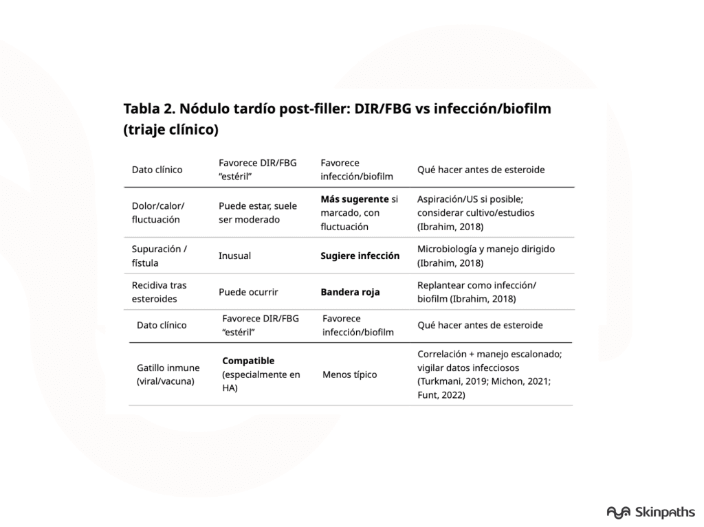
- Erythema, pain, heat, or swelling warrant consideration of inflammatory bowel disease ( IBD) versus infection (including biofilm); this distinction determines whether to prioritize anti-inflammatory treatment or antibiotic/aspiration/culture. Ibrahim, 2018 ; Funt, 2022
- Persistent periorbital edema / Tyndall’s edema (especially in HA)
- Chronic periorbital edema and the Tyndall effect are characteristic complications of tear-trough hyaluronic acid (HTA), influenced by the local plane/anatomy and gel behavior. Vasquez, 2019 ; Skippen, 2020
- Ulceration/necrosis (suspected vascular event)
- The pattern of disproportionate pain, livedo, or necrosis requires urgent, protocol-based management, particularly in HA. Murray, 2021a
Essential information in the medical record: 1- Product (brand if known), dates, site/plane, retreatment. 2- Symptoms: pain, warmth, fluctuation, fever, discharge, recurrences. 3- Triggers: recent systemic infection, vaccination, dental procedures. Turkmani, 2019 ; Michon, 2021 ; Ibrahim, 2018
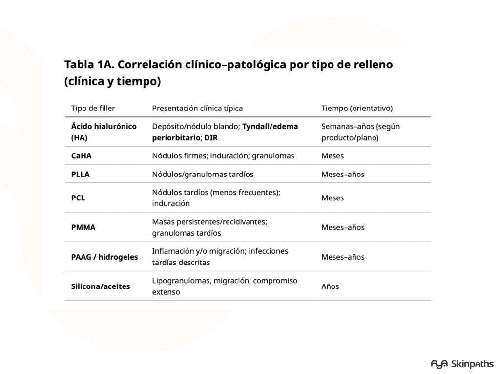
Histological patterns: the «traces» of the material
In clinicopathological series, in addition to the classic granuloma, mixed patterns are described with fibrosis, pseudocavities, chronic inflammation, and, when applicable, suppurative or infectious granulomatous patterns. El-Khalawany, 2015 ; Lee, 2015
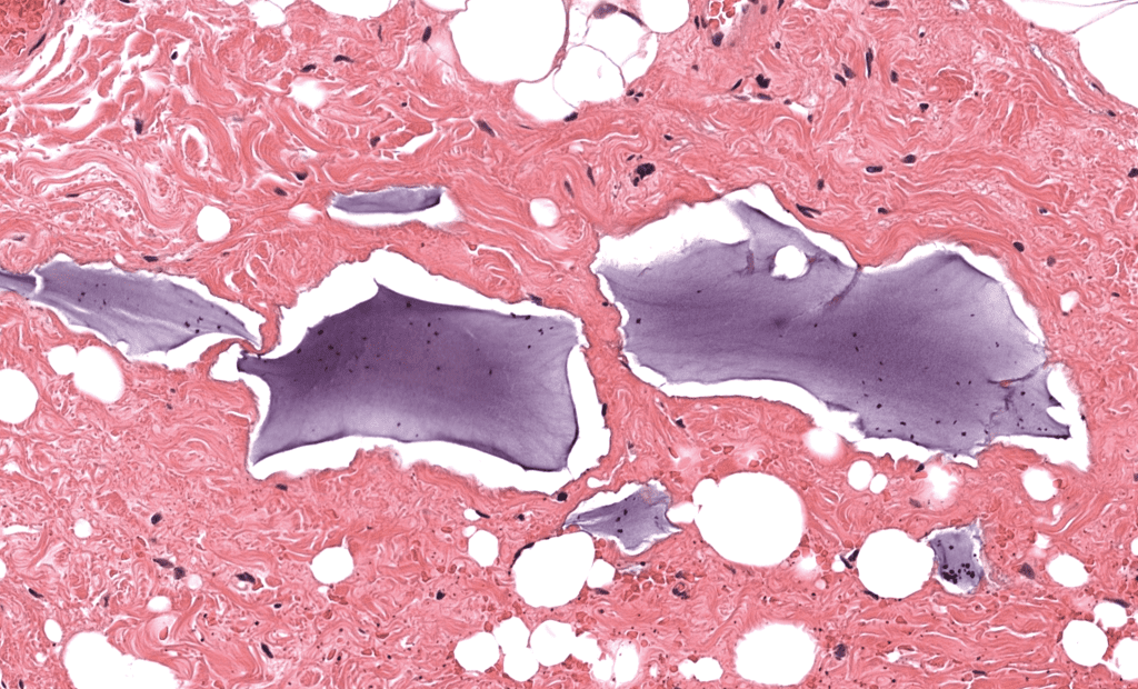
Morphological “signatures” (H&E + simple tricks)
- Hyaluronic acid (HA): amorphous-basophilic deposits resembling “lakes/plaques” or pseudocavities, with variable (sometimes minimal) reaction. El-Khalawany, 2015 ; Bhojani-Lynch, 2017
- CaHA: particulate/microspherular material with variable foreign body response; confirmation of the calcium component can be supported by specific stains. Pavicic, 2013 ; Kadouch, 2017
- PLLA: granulomas with particulate matter and polarization birefringence (a practical finding of high value). Haneke, 2015 ; Storer, 2016
- PMMA: non-degradable, rounded microspheres with late granulomas and a tendency to persist/recurrence. Gelfer, 2007 ; Lemperle, 2009a
- PAAG/hydrogels: amorphous gel with variable reaction; late complications including inflammation, migration, and infection have been described. Ono, 2010 ; Manafi, 2010
- Silicone/oils: “Swiss cheese” vacuolar pattern with histiocytes and giant cells; may be extensive and migratory. Schwartzfarb, 2008 ; Ellis, 2012
Laboratory pitfalls: 1- The material can be altered during processing, leaving «clefts» or spaces; the void may be a clue, not an absence of filler. Lemperle, 2003 2- A targeted approach is better: polarization + 1–2 stains as suspected, instead of indiscriminate panels. El-Khalawany, 2015
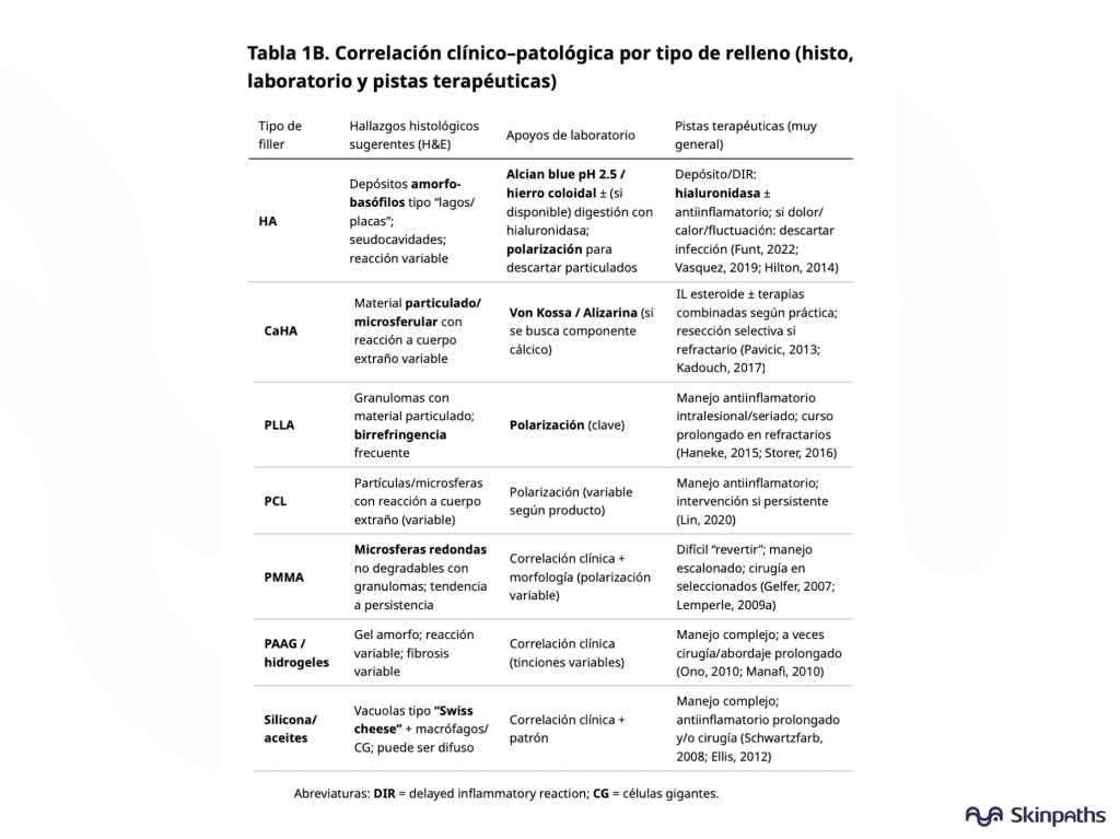
Treatment: scenario-based approach
- Scenario A: Non-inflammatory deposit/nodule
- In HA , hyaluronidase is a key tool for reversing deposition and some local complications (with appropriate technique and dosage). Murray, 2021b ; Hilton, 2014
- In non-HA fillings, management depends on the material and location; it can range from observation to targeted intervention. Haneke, 2015
- Scenario B: DIR/FBG with no infection data
- In HA , there are stepwise algorithms (including hyaluronidase and anti-inflammatory drugs) with close reassessment. Funt, 2022 ; Artzi, 2020
- In biostimulators/permanent implants, prolonged treatment regimens may be required, and in refractory cases, interventional approaches may be necessary. Haneke, 2015 ; Lemperle, 2009b
- Scenario C: Suspected infection/biofilm
- Marked pain, fluctuation, suppuration, fever, or relapse after steroid treatment should prompt aspiration/medical studies and targeted antibiotic therapy, avoiding «encapsulating» the condition with steroids without ruling out infection. Ibrahim, 2018
- Scenario D: Vascular occlusion due to HA (medical emergency)
- Follow guidelines for early recognition and standardized emergency management. Murray, 2021a
Practical algorithm: late post-filling nodule
- Inflammatory?
- No → deposit/irregularity → if HA, consider hyaluronidase; if no-HA, conservative or focal management depending on the material. Haneke, 2015 ; Murray, 2021b
- Yes → next
- Does it suggest infection? (fluctuation, discharge, fever, recurrence, worsens with steroid)
- Yes → aspiration/studies ± antibiotic; biopsy if solid mass or doubt. Ibrahim, 2018
- Not clear → treat as stepwise DIR/FBG and reassess; in HA, hyaluronidase reversal is usually the centerpiece. Funt, 2022
- Persistent/recurrent → confirm material (polarization/targeted staining), reassess infection, and consider interventional management in permanent lesions. El-Khalawany, 2015 ; Lemperle, 2009b
Special section: periorbital involvement
Chronic lower eyelid edema following hyaluronidase injection in the nasojugal sulcus is a well-described phenotype with specific therapeutic approaches (including hyaluronidase in selected cases). Vasquez, 2019 ; Hilton, 2014 ; Skippen, 2020
In conclusion, adverse reactions to dermal fillers are an increasingly common reason for specialist consultation and, at the same time, a diagnostic challenge due to the clinical overlap between deposition , delayed inflammatory reaction (DIR)/foreign body granuloma , and infection (including biofilm formation) . A systematic approach that integrates filler type , chronology , clinical phenotype , and histological pattern allows for a narrower differential diagnosis and guides safer and more effective management. In this context, dermatopathology provides tangible value by recognizing morphological «fingerprints» of the material and selecting high-throughput complementary studies (polarization, targeted staining), thus avoiding both over-treatment with anti-inflammatory agents in potentially infectious cases and unnecessary interventions in deposition nodules. Finally, standardizing information in the requisition (product, dates, plan, symptoms and triggers) and using stepped therapeutic algorithms—with special emphasis on HA, where reversal measures exist—improves results, reduces recurrences and optimizes clinicopathological communication.
Categories: Practicing physicians

Content reviewed by
Dr. Rodolfo Suárez
Médico Patólogo y Dermatólogo Master en Patología Cutánea Avanzada
- Published
- September 18, 2026
- Updated
- September 23, 2026
Medically reviewed by specialists
Our content is based on current scientific evidence and clinical protocols.