
Before reading : This article is intended exclusively for healthcare professionals (physicians, residents, and other professionals with medical training). The content is for educational and academic updating purposes and does not replace individual clinical assessment, professional judgment, or specialized medical consultation . Diagnostic and therapeutic decisions should always be made within the context of each individual patient and under the responsibility of the treating physician.
The information presented is based on concepts widely described in current scientific literature, clinical guidelines, and articles published in specialized medical journals . However, medicine is a constantly evolving field, so the content presented here should not be interpreted as absolute truth or a universal recommendation applicable to all cases . The reader should consider this information as a general guideline and is always advised to review the original sources cited , as well as the most recent available evidence, before making specific clinical decisions.
Key messages
-
Direct immunofluorescence (DIF) is critically dependent on site selection and handling/transport ; errors in these steps decrease yield and generate false negatives. Robinson 2023
-
In autoimmune blistering diseases, the sample for direct immunofluorescence (DIF) is usually perilesional (skin or nearby mucosa, clinically not eroded), while the biopsy for hematoxylin and eosin (H&E) staining is taken from the edge of the blister/lesion . (Witte 2018, Robinson 2023)
-
In small vessel vasculitis, direct immunofluorescence (DIF) is most effective when a very recent lesion is biopsied (ideally within 24–48 hours in practice). Beyond this time, the likelihood of detectable deposits decreases. (Fraticelli 2021, Azanza 2020, Nandeesh 2013)
-
For transport, Michel’s medium is the usual standard; alternatives such as physiological saline may be acceptable only if validated by the laboratory and if delivery is rapid (less than 24 h), with experimental evidence of diagnostic performance under controlled conditions. Vodegel 2004 Robinson 2023
-
Accidental formalinization (fixation in formalin) can render IFD uninterpretable, especially in pemphigus; even brief exposures can erase diagnostic markers and/or induce artifacts (e.g., nuclear fluorochrome). Arbesman 2011
Indications and patient selection
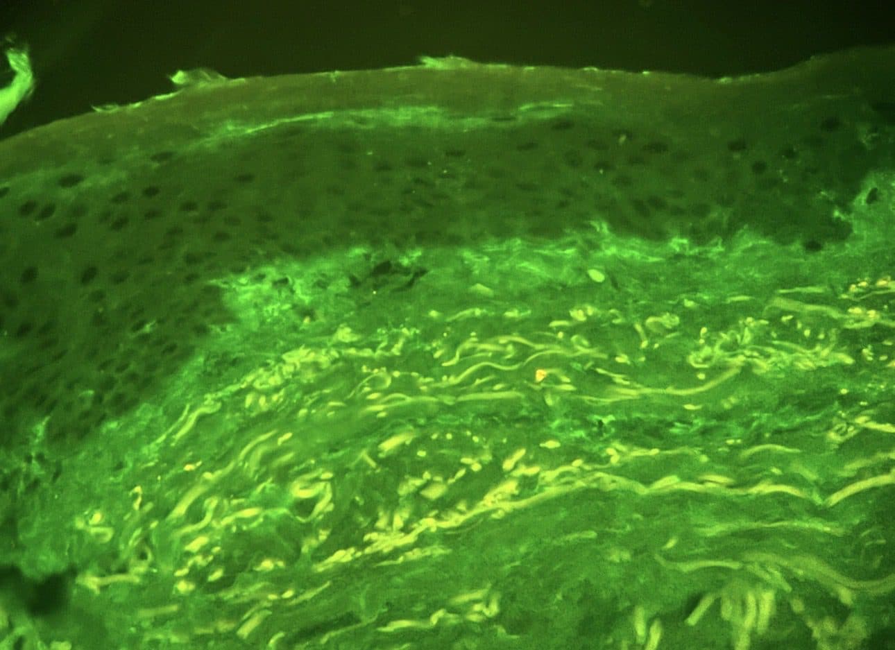
Direct immunofluorescence (DIF) is routinely used when the diagnosis depends on in situ tissue deposits (immunoglobulins/complement) or when clinicopathological correlation requires direct immunopathology, notably in autoimmune blistering diseases (pemphigus/pemphigoid, dermatitis herpetiformis), cutaneous vasculitis, and integration in connective tissue dermatoses (e.g., lupus). Robinson 2023 Schmidt 2010
IFD should be planned as part of a diagnostic package (at least one biopsy for H&E and another for IFD), because the final interpretation often depends on the correlation between immunopathological pattern, histology, and clinical presentation. Robinson 2023, van Beek 2024
In pemphigus/pemphigoid and mucosal pemphigoid, guidelines and consensus statements support direct immunofluorescence (DIF) as a central diagnostic tool; therefore, its omission or inadequate administration delays therapeutic decisions. Schmidt 2015 Rashid 2021 Schmidt 2021
Technique / protocol
1) Preparation (pre-analytical) before infiltrating anesthesia
Coordinate with the laboratory regarding the containers, conditions, and transport methods they use (Michel, physiological saline, or another validated medium), because interlaboratory variability exists and affects the validity of the result. Robinson 2023 Phiske 2026
Plan two samples (H&E + IFD) from the outset and label them unambiguously to avoid the critical error of fixing the sample intended for IFD in formalin. Robinson 2023 Arbesman 2011
Size and depth: A punch of approximately 4 mm is a common recommendation for IFD; if a shave is performed, it should be deep enough when the diagnostic question involves the dermoepidermal junction/vessels. Mahmood 2024, Shetty 2017
2) Site selection based on clinical suspicion (evidence-based practical rules)
A) Autoimmune bullous diseases (pemphigus/pemphigoid, linear IgA, dermatitis herpetiformis)
For IFD, select perilesional skin ≤1 cm from a blister/erosion, avoiding the eroded or frankly necrotic center because this may decrease the detectable deposit. Witte 2018 Robinson 2023
In dermatitis herpetiformis, direct immunofluorescence (DIF) of undamaged perilesional skin is a key diagnostic step, and its effectiveness depends on selecting the correct site. Dmochowski 2019
For H&E biopsy, take a blister margin (including adjacent intact skin) or a complete small lesion, because this defines the level of cleavage and infiltrate. Robinson 2023 Murrell 2020
Anatomical location (classic pitfall in BP): Literature suggests site variation and a risk of false negatives in the lower extremities in some scenarios, but also data showing similar intensities between the trunk and legs in generalized BP; in practice, prioritize the “typical” perilesional area and avoid areas with marked ulceration/stasis when possible. Robinson 2023, Weigand 1989, Anstey 1990
B) Cutaneous vasculitis of small vessels (including IgA vasculitis)
For IFD, choosing the center of a recent injury is preferable because positivity is associated with the time elapsed since the injury and tends to decrease with delay; multiple sources place the practical target at 24–48 h from the onset of the injury outbreak. Fraticelli 2021 Azanza 2020 Robinson 2023
For H&E, a more “established” lesion is often useful to better capture morphological features (the strategy of using two lesions of different ages is used in clinical practice). Robinson 2023 Elston 2016
The usefulness of direct immunofluorescence (DIF) in vasculitis is greatest when integrated with clinical and histological findings, since vascular deposits can be observed in non-vasculitic conditions and require clinicopathological correlation. Nandeesh 2013
C) Lupus erythematosus and “lupus band” (LBT)
In active skin lesions (e.g., cutaneous lupus), IFD biopsy is usually taken from actively lesional skin to demonstrate dermoepidermal junction deposits and/or associated patterns. Robinson 2023 Monroe 1977
When the question is whether a low-dose biopsy (LBT) on non-lesional skin can be used to support systemic lupus diagnosis, there is evidence of differences based on photoexposure and the potential for positivity in photoexposed healthy skin. Therefore, photoprotected skin is preferred if the goal is to maximize specificity. (Maolcatha 2023, Cardinali 1999, Fabré 1991)
D) Oral mucosa and mucosal pemphigoid (MMP) / oral pemphigus vulgaris (OPV)
Direct immunofluorescence (DIF) of mucosal biopsy is recommended for the diagnosis of MMPs, and in cases of negative DIF with high suspicion, sequential biopsies of other sites (additional mucosa and/or skin) may be required to increase diagnostic yield. Rashid 2021 Schmidt 2021
In practice, the “perilesional mucosa” can be challenging due to its fragility; large studies have shown that a biopsy of non-lesional (normal) buccal mucosa can be as sensitive as a perilesional biopsy for MMPs, and in oral PV, the evidence is more nuanced (some data favor perilesional). Carey 2020, Kamaguchi 2018, Mahmood 2024
3) Sample collection (technical points that most impact the IFD reading)
Avoid artifacts caused by crushing (traumatic forceps), desiccation, and cauterization in the fragment intended for IFD, as these degrade the architecture and can compromise pattern interpretation. Elston 2016 Phiske 2026
Immediately separate each cylinder into its correct container and clearly label it “IFD/DIF” to minimize chain of custody errors (the most frequent practical cause of “impossible” IFD). Robinson 2023 Arbesman 2011
4) Transport and preservation
Michel’s medium: It is widely established as a transport medium for preserving tissue immunoreactants prior to direct immunofluorescence (DIF), and there is experimental evidence for the preservation of dermoepidermal junction components for extended periods (up to 28 days in certain contexts), supporting its use when logistics prevent immediate freezing. Woollons 1999 ; Vaughn Jones 1995
Physiological saline (0.9% NaCl): Evidence from paired biopsies of autoimmune/immune complex dermatoses suggests that transport in serum for approximately 24 hours may be adequate under controlled conditions; outside of this setting, it should only be used if validated by the laboratory and delivery is rapid. Vodegel 2004 Robinson 2023
Formalin: It is not an acceptable medium for conventional IFD, and brief exposures can eliminate diagnostic markers (especially in pemphigus) or induce confounding artifactual patterns (e.g., nuclear fluorochrome). Arbesman 2011
5) Minimum clinical information in the application
Always include: primary and differential clinical suspicion, exact location of the site ( and whether it is perilesional/lesional), age of the lesion (in vasculitis), recent treatments (e.g., immunosuppressants), and whether a parallel sample was taken for H&E and/or serology. Robinson 2023 van Beek 2024
Complications + behavior
Punch biopsy in dermatology has a low complication rate, with postoperative bleeding being the most common and infection or vasovagal response occurring less frequently. The risk increases with patient and anatomical site factors, therefore informed consent and careful hemostasis remain mandatory. Yasui 2023 Abhishek 2015
In oral mucosa, clinical series describe a low frequency of major complications and bleeding is usually controlled with pressure, but the operator must avoid neurovascular structures and adjust the depth/size to the local anatomy. Eisen 1992 Carey 2020
Follow-up
The clinical utility of direct immunofluorescence (DIF) increases when the report is interpreted in conjunction with histology and serology (ELISA/IIF depending on availability), and when it is discussed directly with dermatopathology in cases of discordant or negative results with high suspicion. van Beek 2024 Fudge 2018
If the IFD is negative but suspicion remains high (e.g., BP), consider re-biopsy, optimizing the perilesional site and transport logistics, because some false negatives are related to suboptimal sampling or a “subthreshold” immune load . Fudge 2018 Robinson 2023
Bibliography and sources
-
Robinson MR. Direct immunofluorescence. J Clin Aesthet Dermatol. 2023 Dec;16(12 Suppl 2):S33-S35. PMID: 38464740 .
-
Phiske MM, et al. Direct immunofluorescence demystified: Essential insights and recent advances for dermatologists. Indian J Dermatol Venereol Leprol. 2026 Mar-Apr;92(2):179-188. doi:10.25259/IJDVL_95_2024. PMID: 39152802 .
-
Witte M, Zillikens D, Schmidt E. Diagnosis of autoimmune blistering diseases. Front Med (Lausanne). 2018 Nov 2;5:296. doi:10.3389/fmed.2018.00296. PMID: 30450358 .
-
van Beek N, Holtsche MM, Atefi I, Olbrich H, Schmitz MJ, Pruessmann J, Vorobyev A, Schmidt E. State-of-the-art diagnosis of autoimmune blistering diseases. Front Immunol. 2024 Jun 6;15:1363032. doi:10.3389/fimmu.2024.1363032. PMID: 38903493 .
-
Schmidt E, Zillikens D. Modern diagnosis of autoimmune blistering skin diseases. Autoimmun Rev. 2010 Dec;10(2):84-89. doi:10.1016/j.autrev.2010.08.007. PMID: 20713186 .
-
Schmidt E, et al. S2k guideline for the diagnosis of pemphigus vulgaris/foliaceus and bullous pemphigoid. J Dtsch Dermatol Ges. 2015 Jul;13(7):713-727. doi:10.1111/ddg.12612. PMID: 26110729 .
-
Murrell DF, et al. Diagnosis and management of pemphigus: Recommendations of an international panel of experts. J Am Acad Dermatol. 2020 Mar;82(3):575-585.e1. doi:10.1016/j.jaad.2018.02.021. PMID: 29438767 .
-
Schmidt E, et al. European Guidelines (S3) on diagnosis and management of mucous membrane pemphigoid, initiated by the European Academy of Dermatology and Venereology – Part II. J Eur Acad Dermatol Venereol. 2021 Oct;35(10):1926-1948. doi:10.1111/jdv.17395. PMID: 34309078 .
-
Hofmann SC, et al. S2k Guideline for the diagnosis and treatment of mucous membrane pemphigoid. J Dtsch Dermatol Ges. 2022 Nov;20(11):1530-1550. doi:10.1111/ddg.14905. PMID: 36354061 .
-
Mahmood MN. Direct immunofluorescence of skin and oral mucosa: Guidelines for selecting the optimal biopsy site. Dermatopathology (Basel). 2024 Jan 19;11(1):52-61. doi:10.3390/dermatopathology11010006. PMID: 38390848 .
-
Elston DM, Stratman EJ, Miller SJ. Skin biopsy: Biopsy issues in specific diseases. J Am Acad Dermatol. 2016 Jan;74(1):1-16; maybe 17-18. doi:10.1016/j.jaad.2015.06.033. PMID: 26702794 .
-
Fraticelli P, Benfaremo D, Gabrielli A. Diagnosis and management of leukocytoclastic vasculitis. Intern Emerg Med. 2021 Jun;16(4):831-841. doi:10.1007/s11739-021-02688-x. PMID: 33713282 .
-
Chango Azanza JJ, Calle Sarmiento PM, Lopetegui Lia N, Alexander SA, Modi V. Leukocytoclastic vasculitis: An early skin biopsy makes a difference. Cureus. 2020 May 1;12(5):e7912. doi:10.7759/cureus.7912. PMID: 32494527 .
-
Nandeesh BN, Tirumalae R. Direct immunofluorescence in cutaneous vasculitis: Experience from a referral hospital in India. Indian J Dermatol. 2013 Jan;58(1):22-25. doi:10.4103/0019-5154.105280. PMID: 23372207 .
-
Dmochowski M, Gornowicz-Porowska J, Bowszyc-Dmochowska M. An update on direct immunofluorescence for diagnosing dermatitis herpetiformis. Postepy Dermatol Allergol. 2019 Dec;36(6):655-658. doi:10.5114/ada.2019.91415. PMID: 31997990 .
-
Arbesman J, Grover R, Helm TN, Beutner EH. Can direct immunofluorescence testing still be accurate if performed on biopsy specimens after brief inadvertent immersion in formalin? J Am Acad Dermatol. 2011 Jul;65(1):106-111. doi:10.1016/j.jaad.2010.06.019. PMID: 21570155 .
-
Vodegel RM, de Jong MCJM, Meijer HJ, Weytingh MB, Pas HH, Jonkman MF. Enhanced diagnostic immunofluorescence using biopsies transported in saline. BMC Dermatol. 2004 Aug 27;4:10. doi:10.1186/1471-5945-4-10. PMID: 15333135 .
-
Woollons A, Holmes GJ, Gratian MJ, Bhogal BS, Black MM. Michel’s medium: A potential alternative to cryoprotection for tissue transport in the investigation of genetic skin disease. Clin Exp Dermatol. 1999 Nov;24(6):487-489. doi:10.1046/j.1365-2230.1999.00540.x. PMID: 10606956 .
-
Vaughn Jones SA, Palmer I, Bhogal BS, Eady RA, Black MM. The use of Michel’s transport medium for immunofluorescence and immunoelectron microscopy in autoimmune bullous diseases. J Cutan Pathol. 1995 Aug;22(4):365-370. doi:10.1111/j.1600-0560.1995.tb01421.x. PMID: 7499578 .
-
Fudge JG, Crawford RI. Bullous pemphigoid: A 10-year study of discordant results on direct immunofluorescence. J Cutan Med Surg. 2018 Sep-Oct;22(5):472-475. doi:10.1177/1203475418773359. PMID: 29681159 .
-
Anstey A, Venning V, Wojnarowska F, Bhogal B, Black MM. Determination of the optimal site for diagnostic biopsy for direct immunofluorescence in bullous pemphigoid. Clin Exp Dermatol. 1990 Nov;15(6):438-441. doi:10.1111/j.1365-2230.1990.tb02139.x. PMID: 2279342 .
-
Weigand DA, Clements MK. Direct immunofluorescence in bullous pemphigoid: Effects of extent and location of lesions. J Am Acad Dermatol. 1989 Mar;20(3):437-440. doi:10.1016/S0190-9622(89)70054-2. PMID: 2645321 .
-
Carey B, Joshi S, Abdelghani A, Mee J, Andiappan M, Setterfield J. The optimal oral biopsy site for diagnosis of mucous membrane pemphigoid and pemphigus vulgaris. Br J Dermatol. 2020 Mar;182(3):747-753. doi:10.1111/bjd.18032. PMID: 31021396 .
-
Kamaguchi M, Iwata H, Ujiie I, Ujiie H, Sato J, Kitagawa Y, Shimizu H. Direct immunofluorescence using non-lesional buccal mucosa in mucous membrane pemphigoid. Front Med (Lausanne). 2018 Feb 8;5:20. doi:10.3389/fmed.2018.00020. PMID: 29473040 .
-
Rashid H, et al. Assessment of diagnostic strategy for mucous membrane pemphigoid. JAMA Dermatol. 2021 Jul 1;157(7):780-787. doi:10.1001/jamadermatol.2021.1036. PMID: 33909024 .
-
Eisen D. The oral mucosal punch biopsy: A report of 140 cases. Arch Dermatol. 1992 Jun;128(6):815-817. PMID: 1599270 .
-
Ní Maolcatha S, Nic Dhonncha E, O’Connor C, Dinneen S, Heffron CCBB. The lupus band test: A review of the sensitivity and specificity in the diagnosis of lupus erythematosus. Skin Health Dis. 2023 Jul 11;3(4):e205. doi:10.1002/ski2.205. PMID: 37538339 .
-
Cardinali C, Caproni M, Fabbri P. The utility of the lupus band test on sun-protected non-lesional skin for the diagnosis of systemic lupus erythematosus. Clin Exp Rheumatol. 1999 Jul-Aug;17(4):427-432. PMID: 10464552 .
-
Fabré VC, Lear S, Reichlin M, Hodge SJ, Callen JP. Twenty percent of biopsy specimens from sun-exposed skin of normal young adults demonstrate positive immunofluorescence. Arch Dermatol. 1991 Jul;127(7):1006-1011. doi:10.1001/archderm.1991.01680060080008. PMID: 2064397 .
-
Monroe EW. Lupus band test. Arch Dermatol. 1977 Jun;113(6):830-834. PMID: 326195 .
-
Shetty VM, Subramaniam K, Rao R. Utility of immunofluorescence in dermatology. Indian Dermatol Online J. 2017 Jan-Feb;8(1):1-8. doi:10.4103/2229-5178.198774. PMID: 28217464 .
-
Yasui Y, et al. Complications and risk factors of punch biopsy: A retrospective large-scale study. J Dermatol. 2023 Jan;50(1):98-101. doi:10.1111/1346-8138.16585. PMID: 36151785 .
-
Abhishek K, Khunger N. Complications of skin biopsy. J Cutan Aesthet Surg. 2015 Oct-Dec;8(4):239-241. doi:10.4103/0974-2077.172206. PMID: 26865792 .
Categories: Practicing physicians

Content reviewed by
Dr. Rodolfo Suárez
Médico Patólogo y Dermatólogo Master en Patología Cutánea Avanzada
- Published
- September 18, 2026
- Updated
- September 23, 2026
Medically reviewed by specialists
Our content is based on current scientific evidence and clinical protocols.