
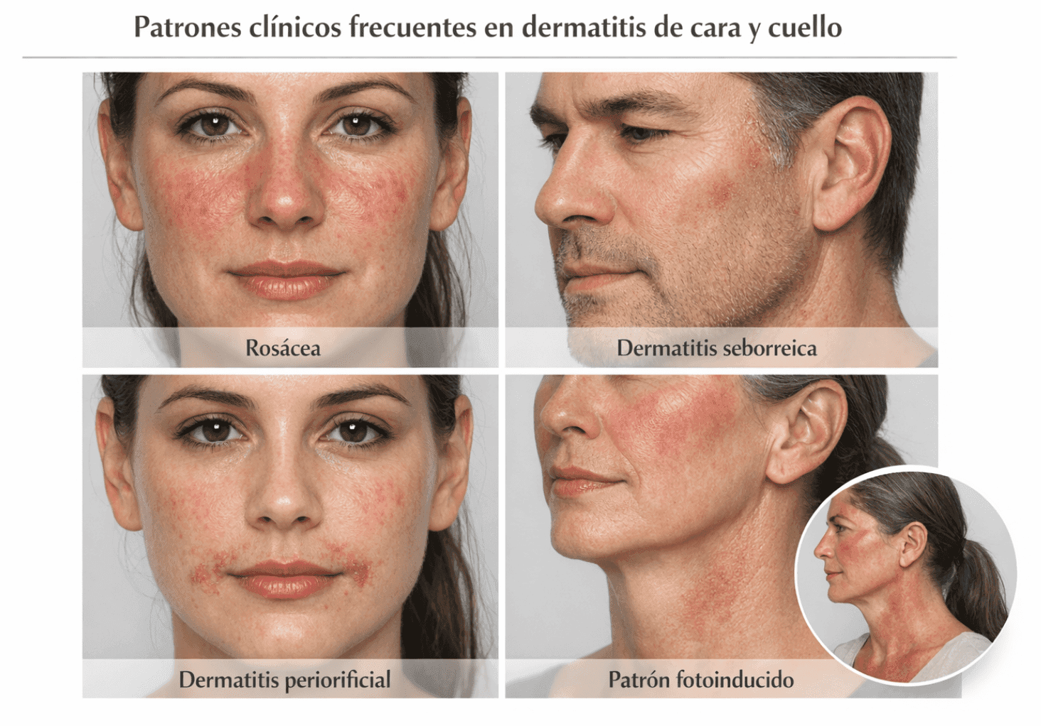
A quick and practical guide to guide a differential diagnosis in facial and neck dermatitis in the consultation, answering 4 key questions.
Before reading: This article is intended exclusively for healthcare professionals (physicians, residents, and other professionals with medical training). The content is for educational and academic updating purposes and does not replace individual clinical assessment, professional judgment, or specialized medical consultation . Diagnostic and therapeutic decisions should always be made within the context of each individual patient and under the responsibility of the treating physician.
The information presented is based on concepts widely described in current scientific literature, clinical guidelines, and articles published in specialized medical journals . However, medicine is a constantly evolving field, so the content presented here should not be interpreted as absolute truth or a universal recommendation applicable to all cases . The reader should consider this information as a general guideline and is always advised to review the original sources cited , as well as the most recent available evidence, before making specific clinical decisions.
1. First of all: 4 quick questions in the consultation
-
Distribution and pattern?
-
Centrofacial? Perioral/periorificial? Eyelids? Lateral/posterior neck
-
Sun-exposed vs. covered? Does it respect nasolabial folds?
-
-
Predominant symptom?
-
Intense itching → eczema (AD / ACD / irritative).
-
Burning/stinging → rosacea, periorificial dermatitis, photodermatosis, irritant.
-
Asymptomatic / very little itching → consider lupus, dermatomyositis, sarcoidosis.
-
-
Patient context?
-
History of atopy , rhinoconjunctivitis, asthma.
-
Chronic use of topical corticosteroids on the face , “active” cosmetics, sunscreens, masks, eye drops.
-
Recent systemic drugs + photoexposure.
-
-
Are there red flags?
-
Fever, weight loss, marked asthenia.
-
Myositis, arthralgia, Raynaud’s phenomenon.
-
Infiltrated nodules or plaques, thick telangiectasias, atrophic scars
-
2. Common clinical patterns and main differential diagnoses
2.1 Centrofacial erythema ± papulo-pustules
Rosacea (erythematotelangiectatic/papulopustular subtype)
-
Clinical keys
-
Persistent central facial erythema (cheeks, nose, chin), telangiectasias.
-
Papule-pustules without comedones .
-
Burning/stinging sensation > itching; flushing with heat, alcohol, stress, spicy foods.
-
-
What helps the differential
-
It does not usually affect retroauricular folds or the scalp like DS.
-
Assess for eye involvement (dry eye, blepharitis).
-
Review previous use of potent topical corticosteroids on the face (steroid rosacea).
-
Facial seborrheic dermatitis (DS)
-
Clinical keys
-
Erythematous-desquamative plaques in nasolabial folds, eyebrows, glabella, retroauricular folds , often scalp and presternal area.
-
Fine-greasy scale; mild-moderate itching.
-
-
Helpful clues
-
Chronic course with flare-ups; association with Malassezia and neurological conditions or HIV.
-
Eyebrow/retroauricular involvement favors DS over rosacea.
-
Head & neck atopic dermatitis (H&N-AD)
-
Clinical keys
-
Patient with history of AD; eczema on face (eyelids, periocular, perioral), neck and upper trunk.
-
Intense itching , generalized xerosis, other typical locations (flexures).
-
-
What guides
-
More “eczema” than flushing; exacerbated by soaps, sweat, friction.
-
It often coexists with Malassezia sensitization and/or superimposed ACD (cosmetics, filters, perfumes).
-
2.2 Perioral / Periorificial Pattern
Periorificial dermatitis (perioral, perinasal, periocular)
-
Clinical keys
-
Perioral and/or perinasal erythematous micropapules and papulopustules, periocular, sparing the vermilion border .
-
Burning/stinging > itching; sometimes a feeling of tightness.
-
-
Typical triggers
-
Topical (or inhaled/nasal) corticosteroids on the face; “heavy” cosmetics, thick mineral filters, fluoridated toothpastes.
-
-
Fast differential
-
Rosacea: more diffuse flushing, telangiectasias, wide centrofacial distribution.
-
AD/ACD: more poorly defined pattern, greater pruritus and lichenification.
-
-
Practical pearl
-
Prioritize discontinuing corticosteroids and triggering cosmetics ; manage with calcineurin inhibitors, metronidazole, macrolides/tetracyclines at anti-inflammatory doses according to severity.
-
ACD perioral / periocular
-
Clinical keys
-
Poorly defined, very pruritic eczema that respects some distance from the mucosa but not as much as the «pure» periorificial eczema.
-
History of eye drops, nail polish, anti-aging creams, sunscreens, face masks.
-
-
To do
-
Think of paraphenylenediamine , fragrances, preservatives, acrylates, eyelash glues, nail polish.
-
2.3 Eyelids and neck
ACD of eyelids and neck (including airborne dermatitis)
-
Clinical keys
-
Very pruritic eyelid eczema (especially upper eyelids), often extending to the malar region/nasojugal sulcus and lateral neck.
-
History of cosmetics, hair dyes, hairsprays, perfumes, filters, necklaces or costume jewelry, workplace exposures.
-
-
Airborne ACD Tracks
-
“Geographic” borders in exposed areas (eyelids, lateral aspect of neck, partially protected submental region).
-
It worsens on weekends in gardening/specific environments, or at work.
-
Irritant dermatitis of the neck/chin
-
Keys
-
Repeated contact with soaps, friction from masks, sweat + athletes, shaving/hair removal.
-
Erythema and scaling are less well-defined; it usually improves with reduced exposure.
-
2.4 Photoinduced pattern (face + V of décolletage + back of neck)
Always consider photoacuteness when: respect of upper eyelids and lower chin, involvement of cheekbones, forehead, nasal root, V of décolletage and back of neck.
Cutaneous lupus erythematosus (acute/subacute)
-
Acute keys (ACLE)
-
Malar rash on nasal wings and cheeks sparing nasolabial folds.
-
It may be mildly pruritic; history of photosensitivity, arthralgia, positive serology.
-
-
Subacute keys (SCLE)
-
Annular/papulosquamous plaques in sun-exposed areas (face, neck, décolleté) with minimal scarring.
-
-
When to perform a biopsy
-
Typical photoinduced pattern + systemic symptoms → biopsy + immunofluorescence + autoimmune panel.
-
Dermatomyositis (DM)
-
Skin clues
-
Heliotrope rash (periorbital erythema/violaceous) + edema; poikiloderma in V of neck and back (signs of “V” and “shawl”).
-
Gottron’s papules on knuckles; variable pruritus.
-
-
Red flags
-
Myositis (difficulty getting up or combing hair), dyspnea, dysphagia, weight loss.
-
In adults, always consider the risk of associated neoplasia.
-
Other photodermatoses to consider
-
PMLE , chronic actinic dermatitis , drug phototoxicity , photocontact (ACD) .
2.5 When “dermatitis” is something else
On the face/neck, certain inflammatory dermatoses can mimic eczema:
-
Cutaneous sarcoidosis : yellowish-pink infiltrated papules/plaques, asymptomatic, sometimes on nasal or periorbital wings.
-
Cutaneous lymphomas / pseudolymphomas : infiltrated plaques, slightly pruritic, chronic course, monochromatic.
-
Facial morphea : indurated, porcelain, depressed plaques, rather asymptomatic.
In any case:
Simple rule : any facial “dermatitis” that is not very itchy, infiltrated or nodular, chronic and asymmetrical deserves a biopsy
3. When to order additional tests
-
Patch tests
-
Almost mandatory in:
-
Chronic eczema of eyelids/neck with no clear cause.
-
Facial “dermatitis” that does not respond to standard AD/DS therapy.
-
H&N-AD phenotype with suspected biologic/JAK-induced dermatosis and possible associated ACD.
-
-
-
Skin biopsy
-
Photoinduced pattern with suspicion of lupus/DM.
-
Infiltrated, asymmetrical, slightly pruritic plaques.
-
Doubt between rosacea vs lupus vs sarcoidosis.
-
-
Laboratory / other
-
ANA, ENA, complement, occult neoplasia profile according to context (lupus/DM).
-
CPK, aldolase, myositis profile, chest imaging if DM is suspected.
-
Annotated bibliography
-
Rozas-Muñoz E, Gamé D, Serra-Baldrich E.
Allergic Contact Dermatitis by Anatomical Regions: Diagnostic Clues. Actas Dermosifiliogr (Engl Ed). 2018;109(6):485-507. DOI: 10.1016/j.ad.2017.05.011. PMID: 29031485. [show_150ms_ease-in] » data-testid=»webpage-citation-pill»> PubMed-
What it’s about: A very practical review of ACD organized by anatomical region (face, eyelids, neck, hands, etc.), with a description of typical clinical patterns and most likely allergens.
-
-
Tolaymat L, Hall MR.
Perioral Dermatitis. In: StatPearls [Internet] . Treasure Island (FL): StatPearls Publishing; 2023–. PMID: 30247843. [show_150ms_ease-in] » data-testid=»webpage-citation-pill»> CENIBiotechnology-
What it’s about: Clinical review chapter on perioral/periorificial dermatitis, including epidemiology, triggering factors (corticosteroids, cosmetics), clinical presentation and treatment.
-
-
Gori N, Ippoliti E, Peris K, Chiricozzi A.
Head and neck atopic dermatitis: still a challenging manifestation in the biological era. Expert Opinion Biol Ther. 2023;23(7):575-577. PMID: 37309908. [show_150ms_ease-in] » data-testid=»webpage-citation-pill»> PubMed-
What it’s about: Expert commentary on the AD phenotype of the head and neck, its high burden on quality of life, and the therapeutic challenges even in the era of biologics.
-
-
Meledathu S, et al.
Update on atopic dermatitis. J Allergy Clin Immunol. 2025; in press at the time of search. [show_150ms_ease-in] » data-testid=»webpage-citation-pill»> ScienceDirect-
What it’s about: A comprehensive and updated review of AD, with emphasis on pathophysiology, clinical phenotypes and modern therapeutic arsenal (biologics and JAKs).
-
-
Farshchian M, et al.
Rosacea. In: StatPearls [Internet] . Treasure Island (FL): StatPearls Publishing; updated 2023. PMID: 32491715. [show_150ms_ease-in] » data-testid=»webpage-citation-pill»> CENIBiotechnology-
What it’s about: Clinical review of rosacea with description of subtypes, typical clinical presentation (flushing, telangiectasias, papules and pustules), triggers and management.
-
-
Nguyen C, et al.
Rosacea: Practical Guidance and Challenges for Clinical Management. Am J Clin Dermatol. 2024;25(1):xx-xx. [show_150ms_ease-in] » data-testid=»webpage-citation-pill»> PMC-
What it’s about: A practical review of rosacea management, including therapeutic algorithms according to subtype and severity.
-
-
Carrizales JP, et al.
The Spectrum of Cutaneous Manifestations in Lupus Erythematosus: A Comprehensive Review. J Clin Med. 2024;13(3). [show_150ms_ease-in] » data-testid=»webpage-citation-pill»> PMC-
What it’s about: A detailed review of the cutaneous forms of lupus (acute, subacute, chronic), with emphasis on clinical patterns and photoinduced distribution.
-
-
Cassard L, et al.
Dermatomyositis: Practical Guidance and Unmet Needs. ImmunoTargets Ther. 2024;13:xx-xx. [show_150ms_ease-in] » data-testid=»webpage-citation-pill»> Dove Medical Press-
What it’s about: A practical review of dermatomyositis, focusing on cutaneous manifestations (heliotrope, V and shawl signs, Gottron’s papules), systemic correlation, and neoplastic risk.
-
-
Gupta AK, et al.
Seborrheic Dermatitis. In: StatPearls [Internet] . Treasure Island (FL): StatPearls Publishing; updated 2024. [show_150ms_ease-in] » data-testid=»webpage-citation-pill»> CENIBiotechnology-
What it’s about: Review on DS, with a focus on clinical aspects, the role of Malassezia, comorbidities (HIV, neurological pathologies) and treatment.
-
Tags
Categories: Featured doctors · Practicing physicians

Content reviewed by
Dr. Rodolfo Suárez
Médico Patólogo y Dermatólogo Master en Patología Cutánea Avanzada
- Published
- September 18, 2026
- Updated
- September 23, 2026
Medically reviewed by specialists
Our content is based on current scientific evidence and clinical protocols.