
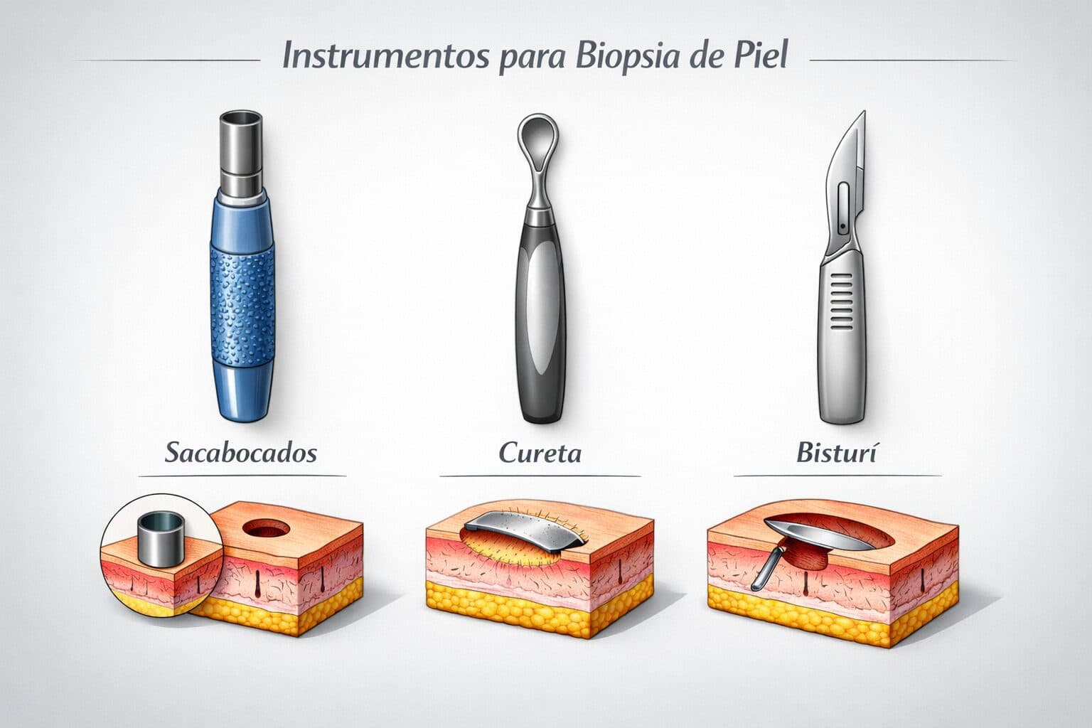
Punch vs. shaved vs. incisional vs. excisional (and what NOT to do in high-risk areas)
Before reading: This article is intended exclusively for healthcare professionals (physicians, residents, and other professionals with medical training). The content is for educational and academic updating purposes and does not replace individual clinical assessment, professional judgment, or specialized medical consultation . Diagnostic and therapeutic decisions should always be made within the context of each individual patient and under the responsibility of the treating physician.
The information presented is based on concepts widely described in current scientific literature, clinical guidelines, and articles published in specialized medical journals . However, medicine is a constantly evolving field, so the content presented here should not be interpreted as absolute truth or a universal recommendation applicable to all cases . The reader should consider this information as a general guideline and is always advised to review the original sources cited , as well as the most recent available evidence, before making specific clinical decisions.
Key messages for the dermatologist
-
More important than «favorite technique» is clinicopathological correlation + site + adequate depth . [show_150ms_ease-in] » data-testid=»webpage-citation-pill»> pubmed.ncbi.nlm.nih.gov
-
In inflammatory conditions, a punch biopsy ≥3–4 mm should be performed , reaching the full dermis (and if panniculitis is suspected, clearly reaching the subcutaneous tissue). [show_150ms_ease-in] » data-testid=»webpage-citation-pill»> pubmed.ncbi.nlm.nih.gov
-
In suspected melanoma, the rule is excisional surgery (when possible), with a narrow margin of 1–3 mm; incisional surgery is reserved for large lesions or in difficult areas. [show_150ms_ease-in] » data-testid=»webpage-citation-pill»> onlinelibrary.wiley.com
-
Face, eyelids, nails, mucous membranes, and genitals are high-risk areas : «easy shaving» here can leave you without a diagnosis or with reconstructive problems. [show_150ms_ease-in] » data-testid=»webpage-citation-pill»> pubmed.ncbi.nlm.nih.gov
-
In cases of alopecia, nail disease, and acral/subungual melanoma, it is crucial to adhere to specific techniques ; an improper biopsy can render the case unrecognizable. [show_150ms_ease-in] » data-testid=»webpage-citation-pill»> pubmed.ncbi.nlm.nih.gov
1. General principles before choosing a technique
- Key questions: Before performing the biopsy:
-
-
What do I want to rule out? (eczema, vasculitis, lymphoma, melanoma, etc.)
-
Where is the expected histological “epicenter”? (epidermis, junction, dermis, panniculus, appendages, nail matrix…)
-
Is the priority diagnosis, margins, or both?
-
Is the area cosmetically critical or functionally complex?
-
- Very practical rules
-
Inflammatory : 3–4 mm punch biopsy of an active, non-excoriated lesion; if bullous, one biopsy of the blister margin for H&E staining and another perilesional biopsy for IFD. [show_150ms_ease-in] » data-testid=»webpage-citation-pill»> pubmed.ncbi.nlm.nih.gov
-
Subcutaneous nodules/panniculitis : 5–6 mm punch or incisional biopsy that reaches the subcutaneous tissue. [show_150ms_ease-in] » data-testid=»webpage-citation-pill»> pubmed.ncbi.nlm.nih.gov
-
Non-pigmented epidermal tumors (small basal cell/squamous cell carcinoma) : deep shave or excisional, depending on size and location. [show_150ms_ease-in] » data-testid=»webpage-citation-pill»> journals.lww.com
-
Atypical pigmented lesions/suspected melanoma : ideally complete excision with narrow margins. Avoid superficial shaves. [show_150ms_ease-in] » data-testid=»webpage-citation-pill»> onlinelibrary.wiley.com
-
In melanoma, the general rule is excisional biopsy (when possible), with narrow margins of 1-3 mm.
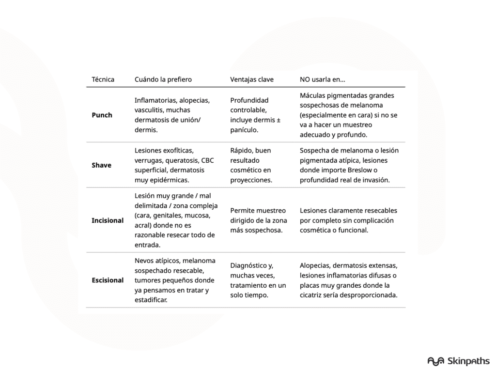
Quick table: advantages and disadvantages according to the technique used
2. Choice according to type of injury
- Inflammatory lesions (“rash”)
- General rule : punch 3–4 mm, over a typical and relatively recent lesion, avoiding:
-
- Areas that are heavily abraded
- Areas that are very chronically lichenized, unless that’s what you want to document.
-
- Vasculitis : purpuric lesion of 24–48 h, deep punch biopsy (dermis + subcutaneous fat if medium-vessel vasculitis is suspected). [show_150ms_ease-in] » data-testid=»webpage-citation-pill»> pubmed.ncbi.nlm.nih.gov
- Bullous disease :
- 1 blister margin biopsy for H&E.
- 1 biopsy on perilesional skin for DIF (not inside the blister) . [show_150ms_ease-in] » data-testid=»webpage-citation-pill»> Frontiers
- General rule : punch 3–4 mm, over a typical and relatively recent lesion, avoiding:
- Non-pigmented tumors
- Small CBC/CEC lesions in trunk and extremities:
- Deep shave (type “saucerization”) or excisional depending on size and therapeutic plan.
- Infiltrative or morpheaform basal cell carcinoma :
- Better incisional or deep excisional that allows evaluation of initial pattern and margins.
- Subcutaneous tumor:
- Large punch (5–6 mm) or fusiform incisional up to panniculus.
- Small CBC/CEC lesions in trunk and extremities:
- Pigmented lesions and melanoma
- Suspected melanoma, <1–1.5 cm and in an accessible area
- Complete excisional resection with a 1–3 mm skin margin. [show_150ms_ease-in] » data-testid=»webpage-citation-pill»> onlinelibrary.wiley.com
- Very large, lentiginous lesion or lesion on the face / acral / genital
- Incisional (elliptic or punch) of the most clinically-dermatoscopically atypical area .
- What NOT to do in suspected melanocytic necrotizing ulcers :
- Superficial shave (risk of underestimating Breslow, margins and architectural pattern).
- Multiple marginal biopsies not including the most doubtful area.
- Suspected melanoma, <1–1.5 cm and in an accessible area
A common mistake to avoid is routinely performing shave biopsies «because it’s faster,» including melanocytic lesions.
3. Choice according to location: “problem” areas and what NOT to do
- Face and eyelids
- What TO do
- Suspicious pigmented lesions (lentigo maligna, facial melanoma):
- Deep incisional or punch aimed at the darker/asymmetrical area.
- basal cell carcinoma / epidermoid:
- Deep or excisional shave, planning the reconstruction from the beginning.
- Inflammatory facial dermatoses:
- Punch 3–4 mm in representative area, avoiding nasolabial folds or free eyelid if another area is available
- What NOT to do
- Superficial shave of a facial macule highly suspicious for melanoma.
- Very small punch (2 mm) in a facial injury where you need full architecture.
- Biopsies that penetrate the free edge of the eyelid are not essential (prefer planned resections with ophthalmic/plastic surgeon).
- What TO do
- Scalp / alopecia
- What TO do
- Cicatricial and non-scarring alopecias:
- One or two 4 mm biopsies; ideally one for cross- sections and one for vertical sections . [show_150ms_ease-in] » data-testid=»webpage-citation-pill»> pubmed.ncbi.nlm.nih.gov
- Take the sample:
- In the area of activity (edge) in scar tissue.
- In less populated areas, in non-scarring areas.
- Scalp dermatosis (psoriasis, eczema, etc.): 3–4 mm punch on active plaque.
- Cicatricial and non-scarring alopecias:
- What NOT to do
- Shaving the scalp when there is diagnostic doubt about alopecia.
- Punch <4 mm for alopecia (you lose follicular counting ability).
- What TO do
- Hands, feet and acral areas
- What TO do
- Pigmented lesions: incisional or excisional always full thickness , including acral ridges and furrows.
- Inflammatory acral hyperkeratosis: deep punch to panniculus (keratodermas, psoriasiform, acral hyperkeratotic eczema).
- What NOT to do
- Superficial shave of pigmented acral macula.
- Tangential biopsies in plants/palms that only take thick stratum corneum.
- What TO do
- Nails (nail apparatus)
- What TO do
- In suspected longitudinal melanonychia :
- Longitudinal or excisional biopsy of the pigmented tract including matrix and bed, with a dorsal or lateral approach. [show_150ms_ease-in] » data-testid=»webpage-citation-pill»> pmc.ncbi.nlm.nih.gov
- In non-pigmented nail dystrophies:
- Biopsy directed at the matrix or bed according to the suspicion (e.g. onychomatricoma vs. onychopapilloma).
- In suspected longitudinal melanonychia :
- What NOT to do
- Shave of the nail plate only (it provides almost nothing except in onychomycosis, and often not even that).
- Blind punch into the matrix without visualizing structures (high risk of permanent longitudinal scarring).
- Biopsy only the hyponychium of a longitudinal melanonychia without including the matrix.
- What TO do
- Oral mucosa and mucous lip
- What TO do
- White/red, erosive or pigmented lesions:
- Incisional with small flap or wedge, including complete epithelium and submucosal band.
- For vesicular-bullous lesions:
- Perilesional mucosal biopsy for DIF; preferably in a non-ulcerated area.
- White/red, erosive or pigmented lesions:
- What NOT to do
- Very superficial shave that leaves only atrophic epithelium and no submucosa.
- Very small biopsies (<3 mm) of lesions suspected of being dysplasia/carcinoma.
- What TO do
- External genitalia and perianal region
- What TO do
- Pigmented vulvar/penile lesions:
- Deep incisional or controlled excisional, attempting to include the most atypical area.
- Chronic inflammatory dermatoses (lichen sclerosus, lichen planus, chronic eczema):
- Punch 3–4 mm, including dermoepidermal junction and some panniculus if possible.
- Pigmented vulvar/penile lesions:
- What NOT to do
- Superficial shave of pigmented vulvar or perianal lesion.
- Very small biopsies that do not allow assessment of architecture (important in VIN, PeIN, melanosis, etc.).
- Clinical information is just as important as choosing the right biopsy technique; don’t forget to write it down on the form!
- What TO do
4. Common mistakes to avoid (mental checklist)
-
Routine shaving of all areas «because it’s faster,» including at-risk melanocytic lesions.
-
Punch too small or superficial in complex inflammatory conditions, alopecia, or panniculitis. [show_150ms_ease-in] » data-testid=»webpage-citation-pill»> pubmed.ncbi.nlm.nih.gov
-
Biopsying heavily treated areas (potent topical steroids, recent cryotherapy) when more «virgin» skin is available. [show_150ms_ease-in] » data-testid=»webpage-citation-pill»> Frontiers
-
Do not indicate on the form the type of injury, duration of the injury, previous treatments, and differential clinical diagnosis. [show_150ms_ease-in] » data-testid=»webpage-citation-pill»> www1.racgp.org.au
-
In alopecia, do not specify whether the laboratory should make cross-sections, vertical sections, or both.
Categories: Practicing physicians

Content reviewed by
Dr. Rodolfo Suárez
Médico Patólogo y Dermatólogo Master en Patología Cutánea Avanzada
- Published
- September 18, 2026
- Updated
- September 23, 2026
Medically reviewed by specialists
Our content is based on current scientific evidence and clinical protocols.