
Before you read: At Skinpaths, we distinguish between opinion and evidence. This content is based on indexed medical literature and recommendations from scientific societies. At the end, you will find a complete bibliography for verification and further study.
Learning objectives
-
To reliably identify the layers of the normal epidermis in histology (standard hematoxylin and eosin staining).
-
Understanding cornification as a protective barrier process (from basal keratinocyte to corneocyte).
-
Recognize the key resident cells (keratinocytes, melanocytes, Langerhans cells, Merkel cells): their location and function.
-
Distinguish normal variations by anatomical site (thick vs thin skin).
-
Avoid frequent reading errors (pitfalls) due to cut orientation and artifacts .
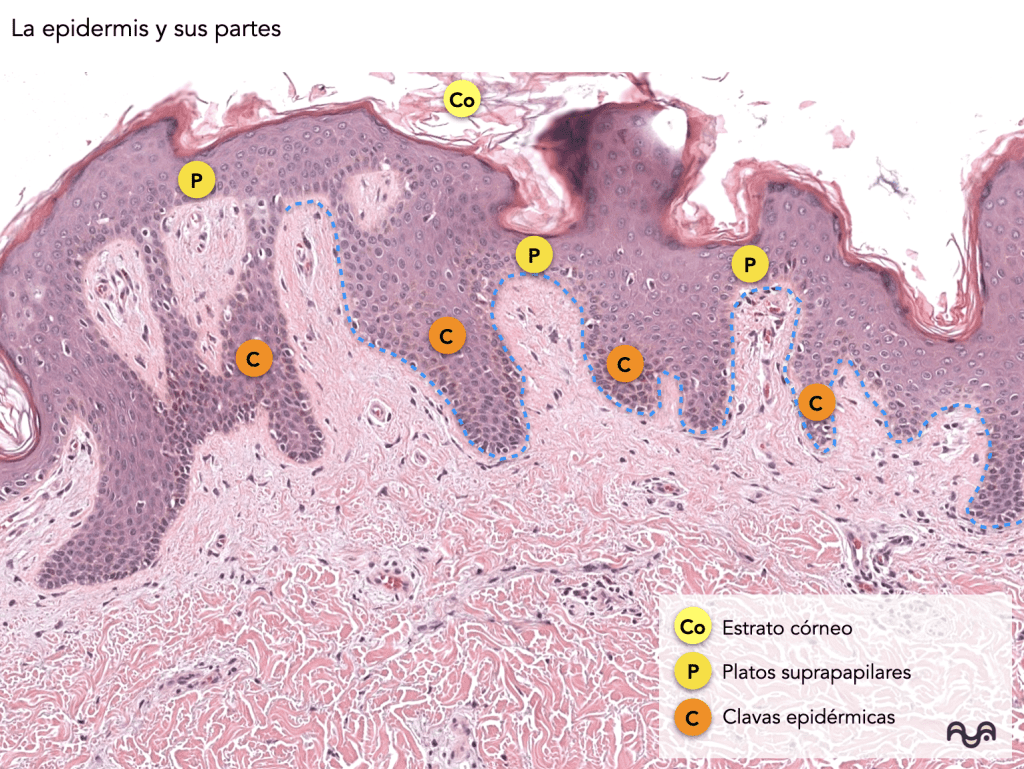
Founding facts (what you absolutely must know)
-
The normal epidermis is a keratinized stratified squamous epithelium and is avascular ; its nutrition depends on diffusion from the dermis. Yousef 2024
-
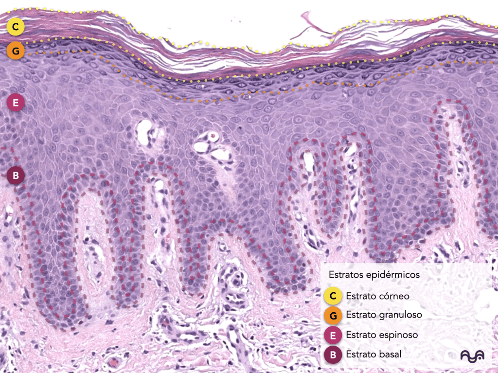
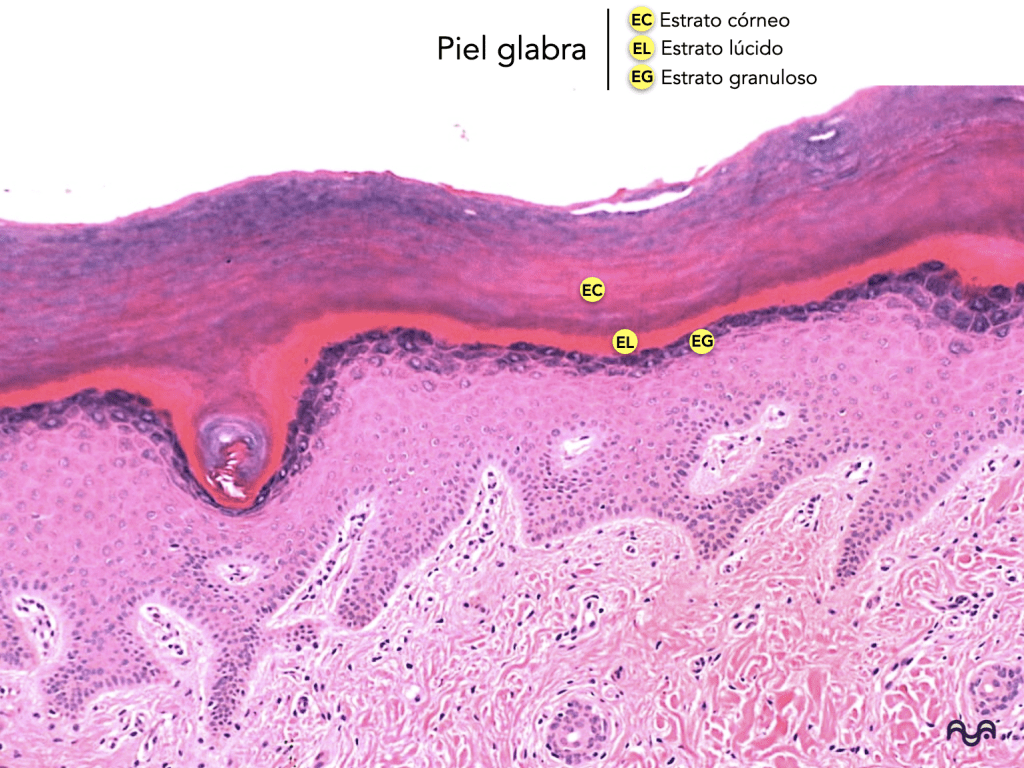
Layers (deep → superficial): basal, spinous, granular, and cornified. The stratum lucidum is observed mainly in thick skin (palms/soles). Yousef 2024
-
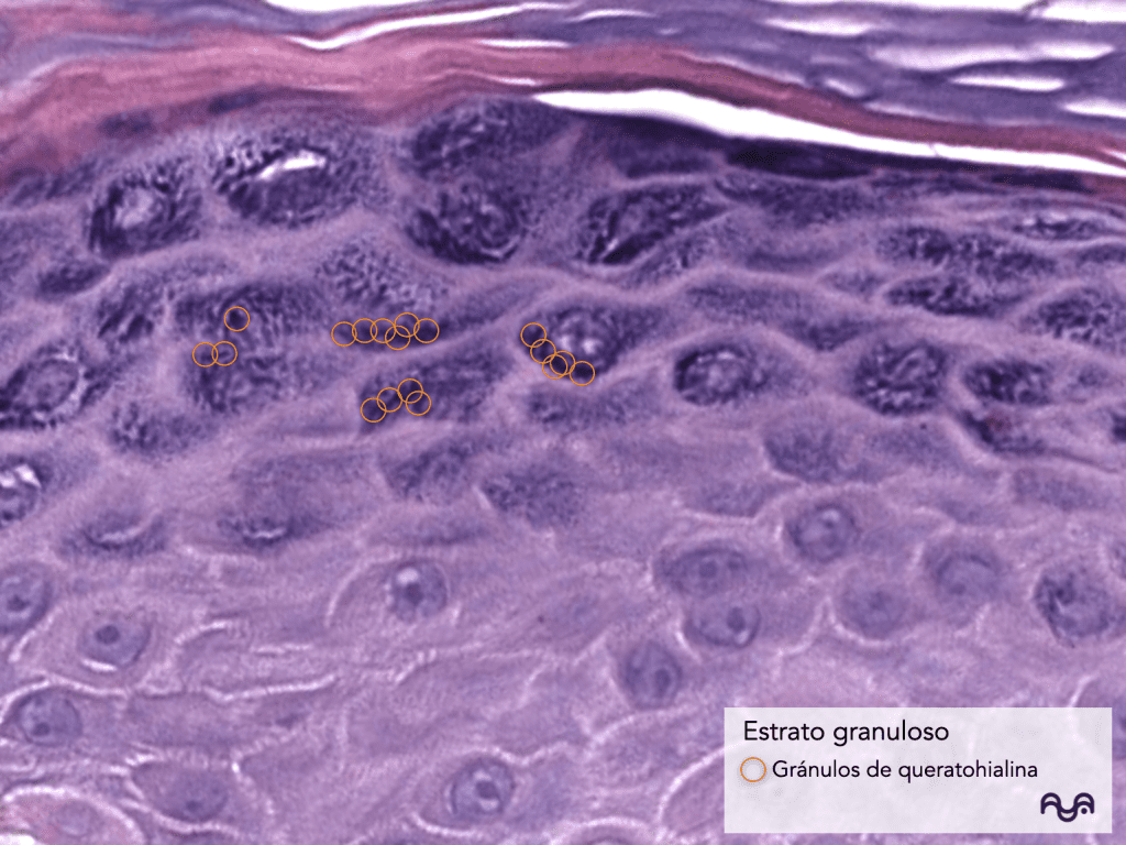
The granular layer is recognized by the presence of basophilic granules (keratohyalin). Yousef 2024
-
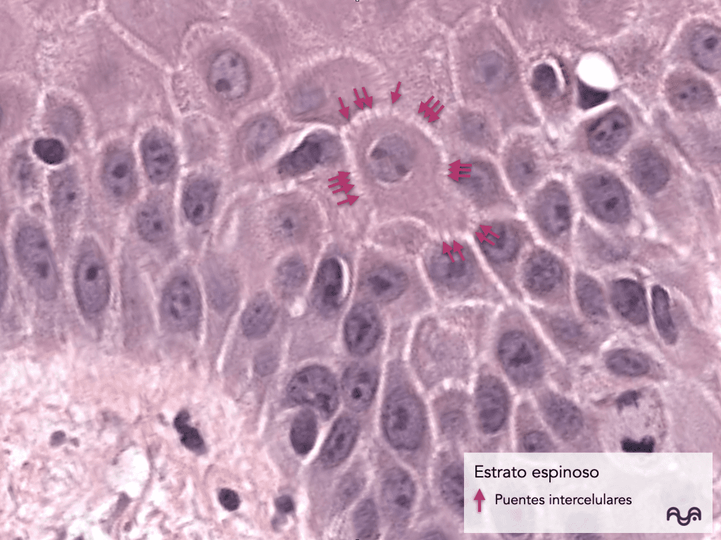
The «spiky» appearance of the stratum spinosum is due to intercellular bridges (desmosomes) that may be highlighted by retraction during processing; this is expected in normal skin. Yousef 2024
-
The stratum corneum functions as a » brick and mortar » barrier: corneocytes + organized extracellular lipids. Del Rosso 2016
-
The epidermal calcium gradient participates in keratinocyte differentiation and in the formation of this barrier. Bikle 2012
-
Filaggrin contributes to the aggregation of keratin filaments and hydration components (NMF) derived from their processing: a key concept for understanding the “normal barrier”. Sandilands 2006
-
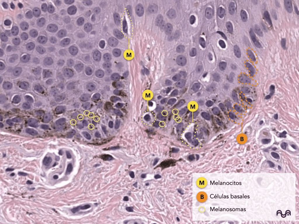
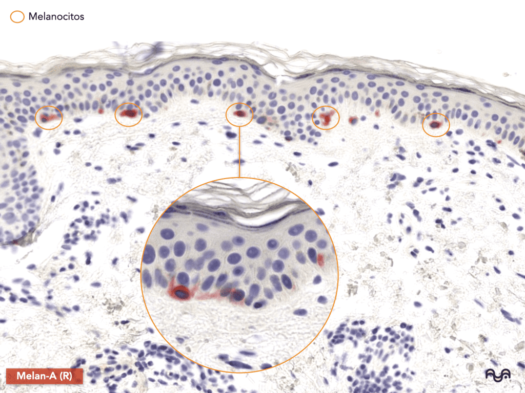
Melanocytes are derived from the neural crest , reside primarily in the basal layer , and transfer melanosomes («packets of melanin») to keratinocytes. Mort 2015
-
Langerhans cells are epidermal immune sentinels; they are not easily identified in normal skin using H&E staining without additional techniques. Romani 2010
-
Merkel cells participate in mechanotransduction in complexes with neurons; they are scarce and do not usually stand out in routine staining (H&E). Woo 2015
-
Thick skin has a thicker epidermis and a prominent stratum corneum; thin skin usually has a thinner epidermis and is associated with the presence of dermal appendages. Yousef 2024
-
A large percentage of «doubts» in normal epidermis are resolved with 3 questions: where is the granular layer?, is the cut perpendicular?, is it thick or thin skin?
Key concepts
Normal epidermis in one sentence
A continuously changing tissue that converts proliferative basal keratinocytes into specialized barrier anucleate corneocytes .
Cornification (terminal keratinization): what does “normal” mean?
In practical terms, “normal” implies:
-
Progressive nuclear loss of the keratinocyte as it reaches the surface.
-
increased structural proteins and envelope formation.
-
extracellular lipid organization that seals the barrier.
Del Rosso 2016 ; Bikle 2012
Microscopic view
“Top-down” method
-
Locate the stratum corneum : the outermost layer.
-
Find the granular layer (locate the basophilic granules): once you see it, the rest falls into place. Yousef 2024
-
Below is the spinous layer : polygonal keratinocytes; apparent intercellular bridges.
-
The basal layer is the deepest row, attached to the dermal stroma (the implantation “line”).
-
Decide on the anatomical site : if the epidermis is very thick and the stratum corneum is dominant, consider palmoplantar skin (possibly lucid). Yousef 2024
Visual pearl: “basket-weave”
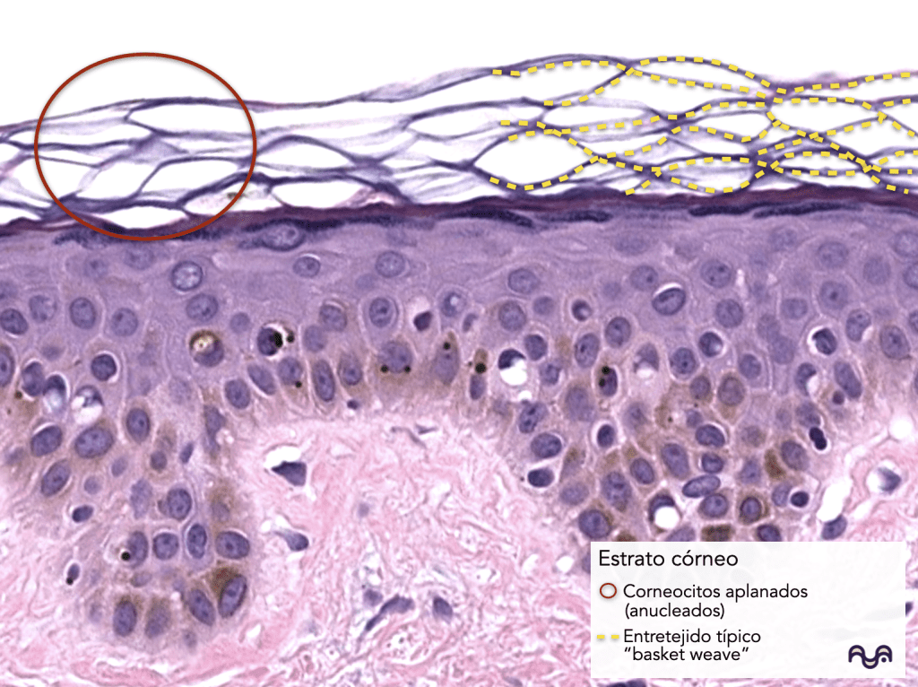
In normal skin (especially thin skin), the stratum corneum may appear with a loose, «basket-like» pattern due to the arrangement of keratin lamellae and processing. This is not a diagnosis; it is a useful morphological description for recognizing normality in many areas.
Microscopic evaluation
Step-by-step approach (90 seconds)
Step 1 — Low magnification (2×–4×): map
-
Is the epidermis thin or thick ?
-
Are there appendages that «dominate» the field and cause you to lose your epidermis?
-
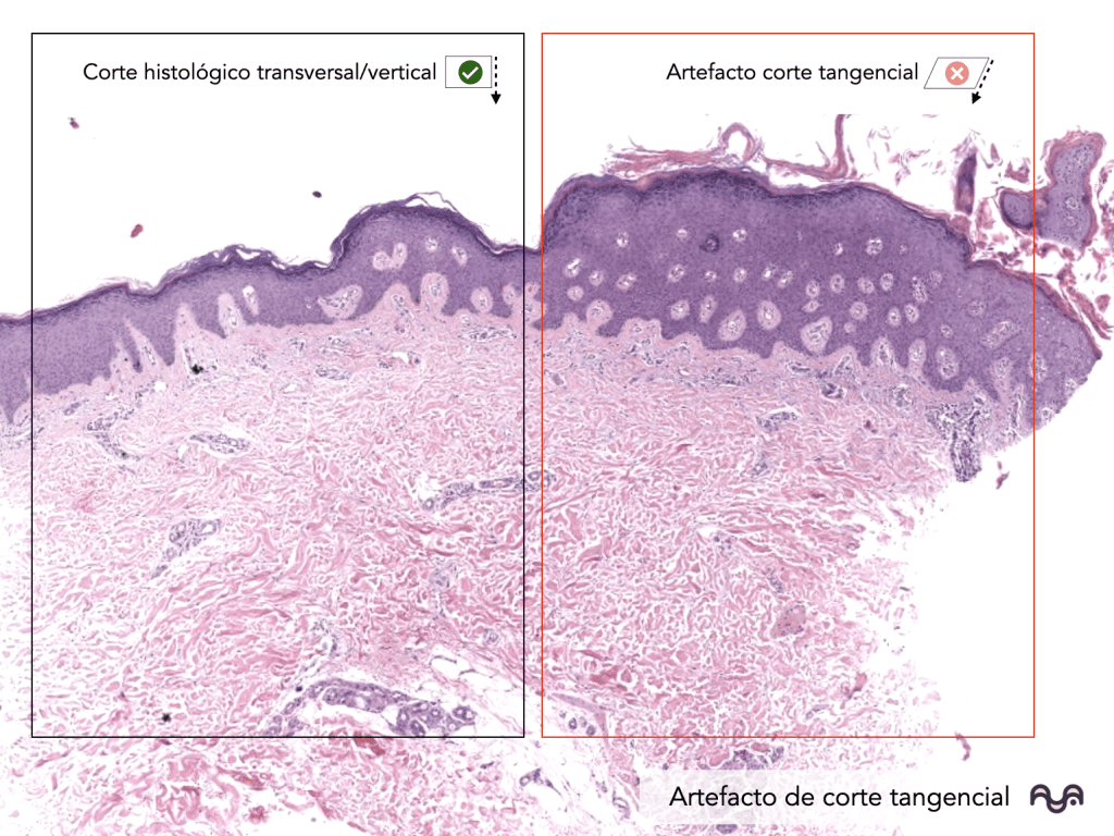
Does the cut appear perpendicular (ideal) or tangential (could confuse evaluation)?
Step 2 — Medium magnification (10×): strata
-
Identify the layers: corneous → granular → spinous → basal .
-
If you don’t find any granular tissue, don’t assume there’s a pathology: it may depend on the location and the cut .
Step 3 — High magnification (20×–40×): cell assessment
-
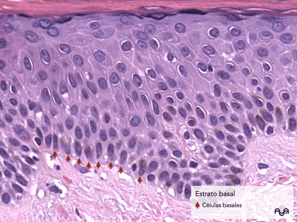
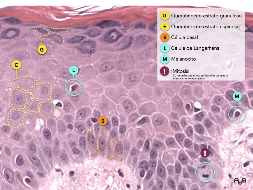
Basal : smaller cells, more basophilic nuclei, and scant cytoplasm. Aligned in a «row».
-
Spiny : polygonal, larger cytoplasm; “bridges” may be prominent.
-
Granular : basophilic granules.
-
Corneal : anucleate eosinophilic laminae.
Strata (what they are, what they do, how to recognize them)
1) Basal (germinative) layer
-
What it is: the deepest layer of keratinocytes, adjacent to the basement membrane.
-
Function: proliferation and maintenance of the epithelium; anchoring to the basal zone (hemidesmosomes as a structural concept). Yousef 2024
-
How to recognize it: a row of more basophilic cells, “orderly” at the dermoepidermal border.
2) Spinous layer
-
What it is: suprabasal layers of maturing keratinocytes.
-
Function: mechanical integrity and cell cohesion.
-
How to recognize it: polygonal keratinocytes; intercellular bridges may be seen (sometimes by retraction of processing).
3) Granular layer
-
What it is: 1–3 layers of «horizontalized» (variable) cells with keratohyalin granules.
-
Function: critical transition to the barrier portion; protein processing (including filaggrin) and preparation for cornification. Sandilands 2006
-
How to recognize it: granular basophilic “belt”.
4) Stratum lucidum (when present)
-
What it is: a pale band between granular and horny, typical of thick skin.
-
Function: related to cornified transition in glabrous skin (classical concept).
-
How to recognize it: a homogeneous, lighter area, if the cut and location allow. Yousef 2024
5) Stratum corneum
-
What it is: anucleated corneocytes in sheets.
-
Function: physical-chemical barrier and hydration, dependent on lipids and structural proteins. Del Rosso 2016
-
How to recognize it: superficial eosinophilic band «wavy» or in «basket weave» (except in specific anatomical areas), usually without nuclei.
Cell types in the epidermis (origin, function, how to see them)
Keratinocytes (the vast majority)
-
Origin: surface ectoderm.
-
Function: They form the barrier and respond physiologically to stress/inflammation signals. Yousef 2024
-
How to see them: they make up almost the entire epidermis in H&E: their morphology changes by layer.
Melanocytes
-
Origin: neural crest.
-
Function: melanin production and melanosome transfer (photoprotection). Died 2015
-
How to see them in H&E: They are often subtle; suggest them by location (basal) rather than by clearly «seeing» them. In teaching, they are best understood with stains/immuno-staining (see photo with Melan-A staining).
Langerhans cells
-
Origin/role: resident dendritic cells; immune surveillance. Romani 2010
-
How to see them in H&E: not reliable in routine; the important thing here is to know that they exist and where they reside (supradial).
-
Historical curiosity: described by Paul Langerhans (19th century), they were initially interpreted differently due to the available techniques; a good example of how morphology depends on methodology. Romani 2010
Merkel cells
-
Role: mechanotransduction in complexes with neurons. Woo 2015
-
How to spot them: rare; they are not usually consistently identifiable in H&E.
Differentials of “normality” (to avoid confusion)
-
Variation by anatomical site : epidermal thickness and prominence of the granulosum/corneal layer change with site and friction. Yousef 2024
-
Variation by orientation : tangential cuts make the epidermis appear thicker or irregular (without actually being so).
Frequent Pitfalls
-
Tangential section : the main enemy; it artificially “thickens” the epidermis and distorts the layers (see next photo)
-
Retraction : may exaggerate intercellular spaces and bridges; this can be seen normally.
-
Compression/knife marks : bands or “wrinkles” in the fabric that simulate changes.
-
Drying or poor fixation : nuclear hyperbasophilia or loss of cytoplasmic detail.
-
Confusing appendages with epidermis : in hairy skin, the follicle may dominate; always return to the «roof» (corneal) and the granular layer to reorient yourself.
Pearls, pitfalls, and common mistakes
-
If you don’t see any granular texture (or if it appears very thick), before worrying, ask: what area is it? Is the cut perpendicular?
-
Melanocytes/Langerhans/Merkel cells: the key in this module is location and function , not «forcing» their identification in routine H&E staining (because they usually require special stains for visualization). Mort 2015 ; Romani 2010 ; Woo 2015
Recommended readings
Essentials (3–6)
-
Epidermis: general architecture and layers — Yousef 2024
-
Stratum corneum barrier (functional framework) — Del Rosso 2016
-
Keratinocyte differentiation and calcium — Bikle 2012
-
Melanocytes: normal development (to understand their discretion in H&E) — Mort 2015
To delve deeper (3–8)
-
Filaggrin and barrier biology — Sandilands 2006
-
Langerhans and cutaneous dendritic cells (integrative view) — Romani 2010
-
Merkel cells and mechanosension — Woo 2015
Bibliography and sources
-
Yousef H, Alhajj M, Fakoya AO, Sharma S. Anatomy, Skin (Integument), Epidermis. In: StatPearls [Internet] . Treasure Island (FL): StatPearls Publishing; 2024. Yousef 2024
-
Del Rosso JQ, Zeichner J, Alexis A, et al. Understanding the epidermal barrier in healthy and compromised skin. J Clin Aesthet Dermatol. 2016;9(4 Suppl 1):S2–S8. Del Rosso 2016
-
Bikle DD, Xie Z, Tu CL. Calcium regulation of keratinocyte differentiation. Expert Rev Endocrinol Metab. 2012;7(4):461–472. Bike 2012
-
Sandilands A, Terron-Kwiatkowski A, Hull PR, et al. Prevalent and rare mutations in the gene encoding filaggrin cause ichthyosis vulgaris and predispose individuals to atopic dermatitis. J Invest Dermatol. 2006;126(8):1770–1775. Sandilands 2006
-
Mort RL, Jackson IJ, Patton EE. The melanocyte lineage in development and disease. Development. 2015;142(4):620–632. Death 2015
-
Romani N, Clausen BE, Stoitzner P. Langerhans cells and more: langerin-expressing dendritic cell subsets in the skin. Immunol Rev 2010;234(1):120–141. Romani 2010
-
Woo SH, Lumpkin EA, Patapoutian A. Merkel cells and neurons keep in touch. Trends Cell Biol. 2015;25(2):74–81. Woo 2015
Categories: Students and residents

Content reviewed by
Dr. Rodolfo Suárez
Médico Patólogo y Dermatólogo Master en Patología Cutánea Avanzada
- Published
- September 18, 2026
- Updated
- September 23, 2026
Medically reviewed by specialists
Our content is based on current scientific evidence and clinical protocols.