

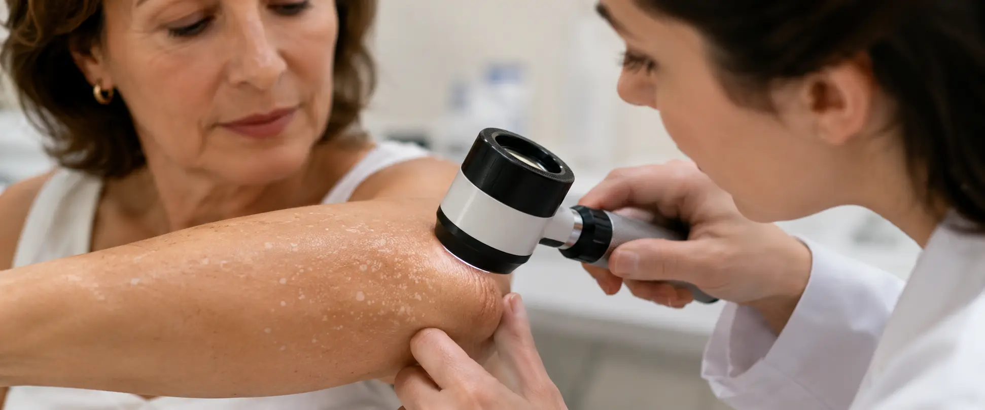
Dermatologists who treat guttate hypomelanosis
Guttate hypomelanosis—also called idiopathic guttate hypomelanosis—is a common cause of small, light-colored or white patches, especially on the arms and legs. In most cases, it is benign, but it can be confused with other conditions that require specific diagnosis, such as vitiligo, pityriasis versicolor, post-inflammatory hypopigmentation, lichen sclerosus, or guttate morphea.
Before reading: En skinpaths We distinguish between opinion and evidence. To write this content, we prioritized indexed medical sources and recommendations from scientific societies. At the end, you will find the complete bibliography for your reference.
Important: This article is for informational purposes only and does not replace medical advice. If you have any questions, persistent symptoms, or your condition worsens, always consult a healthcare professional.
Summary in 60 seconds
- Guttate hypomelanosis produces small, flat, white patches that are usually asymptomatic.
- It is usually seen in areas exposed to the sun, such as forearms and legs, especially on the shins.
- It becomes more frequent with age and may be more noticeable in brown or dark skin due to the contrast.
- It does not usually hurt, itch or peel; when symptoms are present, it is advisable to check if it is another condition.
- It is not an infection and is not considered a precancerous lesion.
- The exact cause is not fully understood; it is associated with skin aging, cumulative sun exposure, genetics, and microtrauma.
- The diagnosis is usually clinical, supported by dermatoscopy if the dermatologist considers it necessary.
- It does not require medical treatment for safety reasons; the available options aim to improve appearance and have variable results.
- Photoprotection helps to care for the skin and reduce the contrast with tanned skin.
- Check if the spots change quickly, bleed, hurt, become inflamed, peel persistently, or appear in unusual areas.
What is guttate hypomelanosis?
Guttate hypomelanosis is an acquired pigmentation disorder. “Hypomelanosis” means a decrease in melanin—the pigment that gives skin its color—and “guttate” refers to small, drop-like lesions. “Idiopathic” indicates that no single, proven cause has been identified.
In practical terms, it manifests as small white or lighter-than-the-surface-skin spots. The classic presentation is round or oval, smooth, flat patches, a few millimeters in size, with relatively well-defined borders. Brown 2025
Typical lesions usually measure 2 to 5 mm, although some may be larger, and are frequently located on the forearms, shins, and other sun-exposed areas. DermNet NZ 2021
In most people, the main impact is cosmetic: the spots can cause concern due to their visibility, especially when they contrast with darker or tanned skin. However, medical confirmation is valuable when the pattern is atypical.
Why does it appear?
Available evidence suggests that guttate hypomelanosis has a multifactorial origin. Proposed factors include skin aging, cumulative ultraviolet exposure, a certain familial predisposition, and repeated minor trauma, although none fully explains all cases. (Juongjin 2016)
Over time, the skin may produce and distribute melanin less evenly. Furthermore, many lesions appear in areas that have been exposed to the sun for decades, such as forearms and legs. This does not mean that every white spot is "from the sun," but rather that cumulative sun exposure seems to contribute in predisposed individuals. PCDS 2021
There are also diseases that can produce light-colored patches and be confused with guttate hypomelanosis. The most relevant are vitiligo, pityriasis versicolor, hypopigmentation following dermatitis or wounds, lichen sclerosus, guttate morphea, and some alterations induced by procedures or medications. The dermatologist not only seeks to "identify" the patch; they also seek to rule out significant mimics. DermNet NZ 2021
What does it look like and what symptoms does it cause?
It is common to observe small, flat, white or whitish spots with no noticeable elevation and no symptoms. They appear most frequently on the shins, forearms, and backs of the limbs; they can also be seen on the trunk, neck, or face, although this is less typical. Brown 2025
They don't usually itch, hurt, or burn. They also don't usually form large, continuous patches. The number of spots may increase with age, and once present, they don't tend to repigment spontaneously in a marked way. DermNet NZ 2021
A helpful clue: if the lesion has fine scaling, itching, redness, rapid growth, crusting, bleeding, or pain, it's best not to assume it's guttate hypomelanosis. These symptoms warrant considering other possibilities.
What can you do today?
The first sensible step is to calmly observe the pattern: approximate size, location, evolution, and symptoms. A well-lit photograph, taken periodically, can help recognize real changes without obsessively checking the skin.
Sun protection is a safe measure. It doesn't necessarily erase existing dark spots, but it protects the skin from ultraviolet damage and can reduce the contrast between tanned and lighter areas. The American Academy of Dermatology recommends broad-spectrum, water-resistant sunscreen with SPF 30 or higher, along with shade and protective clothing. AAD 2026
It can also help to keep your skin well-hydrated, avoid excessive rubbing, and discontinue harsh exfoliation if you notice irritation. For some people, cosmetic camouflage or self-tanners can improve the appearance without subjecting the skin to procedures.
If you're seeking treatment, it's best to do so with a confirmed diagnosis. Topical options and procedures exist—retinoids, calcineurin inhibitors, peels, laser, cryotherapy, dermabrasion, or microneedling in specific contexts—but results vary, and irritation, hyperpigmentation, or unwanted pigment changes may occur.
A recent systematic review found potentially useful interventions, although with methodological limitations and a lack of robust long-term follow-up data. Aravindakshan 2025
What NOT to do?
It is not advisable to apply lemon, vinegar, hydrogen peroxide, baking soda, homemade "depigmenting" agents, or irritating mixtures. There is no evidence that these measures repigment guttate hypomelanosis, and they can cause dermatitis, chemical burns, or post-inflammatory hyperpigmentation.
It is also unwise to use corticosteroids, hydroquinone, strong retinoids, or acids without medical advice. Some of these substances can irritate or thin the skin, cause staining, or worsen contrast if used incorrectly.
Avoid tanning to "even out" your skin tone. Tanning can darken the surrounding healthy skin and make dark spots more noticeable, as well as increase cumulative sun damage. The Skin Cancer Foundation emphasizes that sunscreen is part of a comprehensive strategy that includes shade, clothing, a hat, and UV-protective sunglasses. Skin Cancer Foundation 2026
When to consult?
Consult a dermatologist if any of these signs appear:
- Spots that grow quickly, change shape, or become very irregular.
- Pain, bleeding, persistent scab, wound that does not heal, or inflammation.
- Intense itching, visible flaking, or redness around the affected area.
- Sudden appearance of many spots in a short time.
- Spots on genitals, mucous membranes, or areas with very thin skin.
- Personal history of skin cancer, immunosuppression, or treatments that alter the immune response.
- Doubt between guttate hypomelanosis, vitiligo, fungal infection, or another pigmentary disease.
- Significant emotional or aesthetic impact, even if the spots appear benign.
The consultation does not imply that something is serious; it implies that the skin deserves a precise evaluation when the condition is not typical or causes concern.
What does a dermatologist usually do?
The dermatologist usually begins with a brief medical history: when the spots appeared, whether they have changed, whether they itch or are scaly, what treatments have been used, and how much sun exposure the area has had. Then they examine the entire skin or the relevant areas.
In many cases, the diagnosis is clinical. Dermatoscopy, a lighted magnifying tool, can be used to help evaluate structures not visible to the naked eye. In guttate hypomelanosis, white areas without a pigment network and suggestive dermatoscopic patterns have been described. Ankad 2015
If scaling is present, the physician may consider testing for fungal infections. If the appearance is atypical, or if there is doubt about vitiligo, lichen sclerosus, morphea, or another condition, a skin biopsy may be indicated. In typical guttate hypomelanosis, a biopsy is not usually necessary; it is reserved for cases where the diagnosis is unclear. Brown 2025
When discussing treatment, the conversation should include realistic expectations: the desired aesthetic outcome, the number of sessions, the risk of post-inflammatory hyperpigmentation, cost, discomfort, recovery time, and the possibility of partial response. The decision should not be based on promises of guaranteed repigmentation.
FAQ
Is guttate hypomelanosis dangerous?
- In its typical form, it is considered a benign condition of primarily cosmetic importance. It is not described as a precancerous lesion. Even so, any patch that changes, becomes painful, bleeds, or does not fit the usual pattern should be checked.
Is it the same as vitiligo?
- No. They may look similar because both conditions produce clear or white areas, but their behavior, distribution, and evaluation are different. Dermatoscopy and clinical examination help to differentiate them; sometimes additional tests are required.
Is it contagious?
- It is not considered contagious. It is not a fungal, bacterial, or viral infection. However, some superficial infections, such as tinea versicolor, can also produce light-colored patches; therefore, it is important to check for scaling or itching.
Does it go away on its own?
- It does not usually repigment spontaneously. In some people, the spots remain stable; in others, they may increase in number over time. This does not necessarily mean it is serious.
Does sunscreen remove blemishes?
- It doesn't usually remove them. Its main role is to protect the skin from ultraviolet damage and help prevent the surrounding skin from tanning further, which can make the spots more visible.
Is there a treatment?
- There are cosmetic treatments with varying responses: topical treatments, peels, laser, cryotherapy, dermabrasion, or microneedling techniques in selected settings. None should be considered universally effective for all patients, and some may cause irritation or pigment changes.
Should I be worried if I have a lot of spots?
- The number of lesions can increase with age and does not necessarily indicate a serious illness. It is advisable to consult a doctor if the change was rapid, if there are symptoms, if the lesions are very different from each other, or if there is diagnostic uncertainty.
Bibliography and sources
- Juntongjin P, Laosakul K. Idiopathic guttate hypomelanosis: a review of its etiology, pathogenesis, findings, and treatments. Am J Clin Dermatol. 2016;17(4):403-411. doi:10.1007/s40257-016-0195-3. Available in: https://pubmed.ncbi.nlm.nih.gov/27206417/
- Brown F, Syed HA, Crane JS. Idiopathic Guttate Hypomelanosis. In: StatPearls [Internet]. Treasure Island (FL): StatPearls Publishing; 2026 Jan-. Updated 2025 Feb 17. Available in: https://www.ncbi.nlm.nih.gov/books/NBK482182/
- Ankad BS, Beergouder SL. Dermoscopic evaluation of idiopathic guttate hypomelanosis: a preliminary observation. Indian Dermatol Online J. 2015;6(3):164-167. doi:10.4103/2229-5178.156383. Available in: https://pmc.ncbi.nlm.nih.gov/articles/PMC4439743/
- Kim SK, Kim EH, Kang HY, Lee ES, Sohn S, Kim YC. Comprehensive understanding of idiopathic guttate hypomelanosis: clinical and histopathological correlation. Int J Dermatol. 2010;49(2):162-166. doi:10.1111/j.1365-4632.2009.04209.x. Available in: https://pubmed.ncbi.nlm.nih.gov/20465639/
- Aravindakshan R, Gopinath H, Kaimal S, Pentapati SS, Marimuthu Y. A systematic review of interventions for idiopathic guttate hypomelanosis. Clin Exp Dermatol. 2025;50(9):1741-1747. doi:10.1093/ced/llaf134. Available in: https://pubmed.ncbi.nlm.nih.gov/40192523/
- DermNet NZ. Idiopathic guttate hypomelanosis. DermNet NZ; updated June 2021. Available in: https://dermnetnz.org/topics/idiopathic-guttate-hypomelanosis
- DermNet NZ. Idiopathic guttate hypomelanosis dermoscopy. DermNet NZ; updated December 2019. Available in: https://dermnetnz.org/topics/idiopathic-guttate-hypomelanosis-dermoscopy
- Primary Care Dermatology Society. Idiopathic guttate hypomelanosis. PCDS; updated August 8, 2021. Available in: https://www.pcds.org.uk/clinical-guidance/idiopathic-guttate-hypomelanosis
- Cleveland Clinic. Idiopathic Guttate Hypomelanosis: Causes & Treatment. Cleveland Clinic; updated January 12, 2026. Available in: https://my.clevelandclinic.org/health/diseases/idiopathic-guttate-hypomelanosis
- American Academy of Dermatology Association. Sunscreen FAQs. AAD. Available in: https://www.aad.org/media/stats-sunscreen
- Skin Cancer Foundation. Sunscreen & Your Skin. Skin Cancer Foundation; updated June 2026. Available in: https://www.skincancer.org/skin-cancer-prevention/sun-protection/sunscreen/

 |
|