

The ABCDEs of melanoma is a visual rule for recognizing signs that a mole or spot may warrant medical evaluation. It’s not for making a definitive diagnosis at home, but it can help you decide more clearly when to request a dermatological checkup.
Before you read: At Skinpaths, we distinguish between opinion and evidence. To write this content, we prioritized indexed medical sources and recommendations from scientific societies. You’ll find the complete bibliography at the end for your reference.
Important: This article is for informational purposes only and does not replace medical advice. If you have any questions, persistent symptoms, or your condition worsens, consult a healthcare professional.
Summary in 60 seconds
- Melanoma is a type of skin cancer that originates in melanocytes, the cells related to skin pigment.
- It can appear on a pre-existing mole, but also on skin that previously appeared normal.
- The ABCDE method helps to check spots and moles in an orderly way: Asymmetry, Border, Color, Diameter and Evolution.
- The most important letter in daily life is usually E for evolution : a new change deserves attention.
- A melanoma can be small; a diameter greater than 6 mm is indicative, but does not rule out smaller lesions.
- The «ugly duckling» also matters: a lesion that looks different from the rest of your moles.
- Checking your skin once a month, in good light and without rushing, can help you recognize changes.
- The palms, soles, nails, scalp, and areas not exposed to the sun should also be checked.
- If you experience bleeding, persistent scabbing, growth, new itching, pain, or a different spot, it is advisable to consult a specialist.
- Diagnostic confirmation, when there is suspicion, usually requires dermatological evaluation and, sometimes, a biopsy.
What is it?
Melanoma is a type of skin cancer that develops from melanocytes. Although it can be linked to exposure to ultraviolet radiation, not all melanomas appear in areas clearly exposed to the sun; they can also occur on nails, palms, soles, mucous membranes, or other less visible areas. NCCN 2021
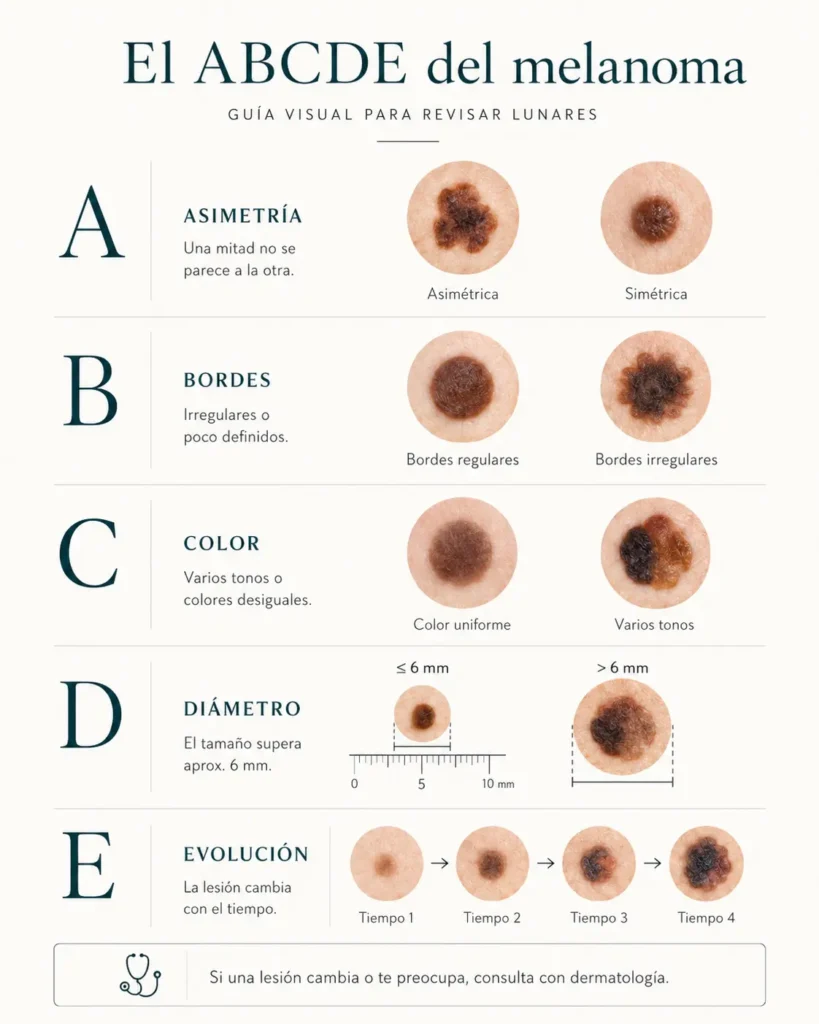
The ABCDE method is an observation tool. Its letters summarize five characteristics that may make a lesion suspicious; any lesion that has at least one of these characteristics requires evaluation by a dermatology specialist.
A (asymmetry)
- Does one half look different from the other?
B (edges)
- Are the edges irregular, blurry, jagged, or poorly defined?
C (color)
- Does it have several shades (2 or more): brown, black, reddish, blue, or uneven areas?
D (diameter)
- Does it measure more than 6 millimeters?
E (evolution)
- Did it change in size, shape, color, thickness, feel, or did it start bleeding?
The American Academy of Dermatology describes these same criteria and notes that melanomas are usually larger than 6 mm at diagnosis, but can be smaller. Therefore, size is helpful, but should not be used as the sole criterion. AAD 2025
Why does melanoma occur?
Melanoma does not have a single cause. In many people, several factors are involved: intense or cumulative sun exposure, sunburns, use of tanning beds, a family or personal history of melanoma, many moles, atypical moles, skin that burns easily, and immunosuppression. The National Cancer Institute also emphasizes that people of any skin color can develop skin cancer. NCI 2025
Evidence supports the idea that ultraviolet radiation is a significant risk factor, but in clinical practice, the message is often reduced to simply “sun exposure.” Some melanomas appear in areas with little sun exposure, and in darker skin tones, they may occur more frequently on the soles of the feet, palms, or under the nails. Skin Cancer Foundation 2025
What does it look like and what symptoms does it cause?
A melanoma can appear as an irregular dark patch, a changing mole, a multi-colored lesion, or a pigmented area that extends beyond the usual borders. It can also appear as a pink, reddish, clear, or skin-colored lesion, which makes some melanomas more difficult to recognize. Skin Cancer Foundation 2025
The American Cancer Society summarizes two especially helpful signs: a new or changing patch, and a lesion that looks different from the others, known as the “ugly duckling” sign. It also recommends consulting a doctor about sores that don’t heal, bleeding, scaling, pain, itching, or changes in texture. American Cancer Society 2025
What can you do today?
The easiest way to apply the ABCDE method is to make it a short, gentle routine, not an anxious quest. Choose one day a month, use good lighting, a full-length mirror, and, if possible, ask for help with your back, scalp, and other problem areas.
A simple and safe method in 5 steps:
- Look at the whole picture. See if your moles look alike. A very different lesion deserves attention.
- Apply the ABCDE rule. You don’t need to find all the letters; a single positive sign justifies consulting a specialist.
- Check forgotten areas. Scalp, ears, lips, neck, armpits, back, buttocks, genitals, palms, soles, between fingers and nails.
- Take a useful photo, not a perfect one. Use the same lighting, the same distance, and a ruler or other size reference. Avoid comparing blurry or filtered photos.
- Decide calmly. If something is new, changes, or worries you, schedule an evaluation. A medical check-up doesn’t mean it’s cancer; it means it deserves an expert opinion.
Important fact: Available scientific evidence supports the educational value of skin self-examination, although its accuracy varies and it does not replace professional evaluation by a dermatologist. Hamidi 2010
What NOT to do?
- It’s not advisable to scratch, cut, burn, squeeze, or try to «remove» a mole at home. It’s also unwise to cover it with makeup for months or wait until it bleeds several times before seeking medical advice.
- Mobile apps and image searches can provide guidance, but they do not replace a dermatological examination.
- An isolated photo can lose relevant information: texture, context of other moles, changes over time, dermatoscopy, and personal risk factors.
- It is also important to avoid the falsely reassuring message: “if it’s small, it doesn’t matter.” Some melanomas can measure less than 6 mm, which is why monitoring their evolution and comparing them to the surrounding skin are crucial. Abbasi 2004
When should I consult?
Consult a dermatologist if any of these signs appear:
- A new mole or spot that grows or attracts attention.
- A mole that changes in size, shape, color, thickness, or texture .
- An injury that meets any letter of the ABCDE .
- A spot that looks different from the rest of your moles: the «ugly duckling».
- Bleeding, persistent scab, wound that does not heal, new pain, tenderness or itching.
- A new dark line on a nail, pigment spreading into the nearby skin, or a progressive change in a nail band.
- New lesions on palms, soles, mucous membranes, or hard-to-see areas.
- Personal or family history of melanoma, many atypical moles, or immunosuppression.
The consultation shouldn’t be seen as a definitive alarm. In dermatology, many suspicious lesions turn out to be benign, but the safest course of action is to have them evaluated promptly by a healthcare professional.
What does a dermatologist usually do?
The dermatologist usually begins with a brief medical history: when the lesion appeared, if it changed, if it bleeds, if it hurts, personal or family history, sunburns, tanning beds, medications, and immunosuppression.
Next, a skin examination is performed. In many cases, dermatoscopy is used, a non-invasive technique that allows the observation of structures that are not easily seen with the naked eye. Systematic reviews have found that dermatoscopy, when used with appropriate training, improves accuracy compared to visual inspection alone. (Dinnes 2018)
If the lesion is suspicious, a biopsy may be recommended. The American Academy of Dermatology guidelines state that the biopsy should yield sufficient histopathological information to confirm the diagnosis and guide management. (Swetter 2019)
Frequently Asked Questions
Are all irregular moles melanoma?
No. Many benign moles can be somewhat asymmetrical or have unusual borders. The ABCDE rule doesn’t diagnose; it helps decide which lesions warrant further investigation.
Is a melanoma always black?
No. It can be brown, black, reddish, pink, bluish, white, or even appear to be the same color as skin. That’s why evolution and the «ugly duckling» are so important.
Does a size greater than 6 mm confirm melanoma?
No. Diameter is a guideline. A lesion larger than 6 mm should be examined closely, but smaller melanomas can also occur. AAD 2025
How often should I check my skin?
For many people, a brief, routine monthly check-up is reasonable. Those with a history of melanoma, numerous moles, atypical moles, or immunosuppression may need a personalized plan with a dermatologist.
What should I do if a mole itches?
Isolated itching does not necessarily mean melanoma. But if the itching is new, persistent, or accompanied by growth, change in color, crusting, bleeding, or pain, it’s advisable to consult a doctor.
Should people with brown or dark skin also apply the ABCDE method?
Yes. Although some risks vary among populations, anyone can develop melanoma. In darker skin, it is especially important to check the soles of the feet, palms, nails, and less exposed areas.
Can I wait and see if it disappears?
It depends on the case, but a pigmented lesion that changes, bleeds, grows, or looks different should not be left unattended for months without evaluation. A prudent wait is different from prolonged delay.
Bibliography and sources
- American Academy of Dermatology Association. What to look for: ABCDEs of melanoma . American Academy of Dermatology; 2025.
- American Cancer Society. Signs and symptoms of melanoma skin cancer . American Cancer Society; 2025.
- Skin Cancer Foundation. Melanoma warning signs and images . Skin Cancer Foundation; 2025.
- National Cancer Institute. Skin Cancer Prevention—PDQ Patient Version . National Cancer Institute; 2025.
- National Comprehensive Cancer Network. NCCN Guidelines for Patients: Melanoma . Version 2.2021. Plymouth Meeting: NCCN Foundation; 2021.
- Abbasi NR, Shaw HM, Rigel DS, Friedman RJ, McCarthy WH, Osman I, et al. Early diagnosis of cutaneous melanoma: revisiting the ABCD criteria . JAMA. 2004;292(22):2771-2776.
- Tsao H, Olazagasti JM, Cordoro KM, Brewer JD, Taylor SC, Bordeaux JS, et al. Early detection of melanoma: reviewing the ABCDEs . J Am Acad Dermatol. 2015;72(4):717-723.
- Swetter SM, Tsao H, Bichakjian CK, Curiel-Lewandrowski C, Elder DE, Gershenwald JE, et al. Guidelines of care for the management of primary cutaneous melanoma . J Am Acad Dermatol. 2019;80(1):208-250.
- Dinnes J, Deeks JJ, Chuchu N, Ferrante di Ruffano L, Matin RN, Thomson DR, et al. Dermoscopy, with and without visual inspection, for diagnosing melanoma in adults . Cochrane Database Syst Rev. 2018;12(12):CD011902.
- Jones OT, Jurascheck LC, van Melle MA, Hickman S, Burrows NP, Hall PN, et al. Dermoscopy for melanoma detection and triage in primary care: a systematic review . BMJ Open. 2019;9(8):e027529.
- Hamidi R, Peng D, Cockburn M. Efficacy of skin self-examination for the early detection of melanoma . Int J Dermatol. 2010;49(2):126-134.
- Weinstock MA, Risica PM, Martin RA, Rakowski W, Smith KJ, Berwick M, et al. Melanoma early detection with thorough skin self-examination: the “Check It Out” randomized trial . Am J Prev Med. 2007;32(6):517-524.
Categories: For everyone · Moles · Skin cancer

Content reviewed by
Dr. Rodolfo Suárez
Médico Patólogo y Dermatólogo Master en Patología Cutánea Avanzada
- Published
- September 18, 2026
- Updated
- September 23, 2026
Medically reviewed by specialists
Our content is based on current scientific evidence and clinical protocols.