

A mole that changes or grows shouldn’t cause panic, but it shouldn’t be ignored either. Many changes have benign causes, but some can be the first sign of skin cancer that should be detected early.
Before you read: This article provides general medical information to help you recognize signs that might warrant a dermatological evaluation. It is not a substitute for a medical consultation and does not allow for online diagnosis. If you have a lesion that changes, bleeds, hurts, grows rapidly, or is accompanied by general symptoms, consult a dermatologist.
What does this sign mean?
A mole that changes or grows is a pigmented lesion that was already on the skin and begins to look different: it increases in size, changes shape, darkens, loses color, becomes irregular, rises, itches, bleeds, or forms crusts.
Not every change means skin cancer. Some moles can change for benign reasons, such as body growth, pregnancy, sun exposure, rubbing, or inflammation. However, a change in a mole is a sign that should be taken seriously, because melanoma can appear as a new lesion or as a pre-existing mole that changes over time . NHS-2023 American-Cancer-Society-2025
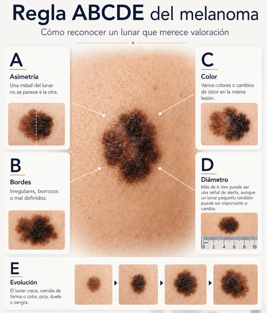
The ABCDE rule is a simple way to remember signs that might make a mole suspicious. A stands for asymmetry: one half doesn’t look like the other. B refers to irregular or poorly defined borders. C indicates changes or mixing of colors, such as brown, black, red, white, or blue. D refers to diameter, especially if it’s larger than 6 mm, although a small mole can also be important if it changes. E stands for evolution: any mole that grows, changes shape, color, or texture, itches, hurts, or bleeds should be evaluated by a dermatologist. This rule doesn’t allow you to diagnose melanoma online, but it helps you recognize when it’s appropriate to consult a doctor. AAD-2026
–
Common and generally less serious causes
In many cases, a lesion that appears to be a changing mole may actually be a benign mole, also called a melanocytic nevus. Moles can appear in childhood or adolescence, and some may change slowly over the years; they can also be altered by sun exposure or pregnancy. DermNet-2023
In children and adolescents, some birthmarks may grow as the body grows. They may also become darker, raised, or develop more hair around puberty. Even so, any irregular, rapid, or unusual change compared to the typical pattern of other moles should be checked. DermNet-2023
Another common cause of confusion is seborrheic keratosis. It is not a true mole, but a benign lesion that usually appears in adulthood. It can be brown, black, rough, or have a «stuck» appearance on the skin. Sometimes it becomes irritated, crusts over, or resembles skin cancer, so dermatoscopy or a biopsy may be required if there is diagnostic uncertainty. DermNet-2023
Changes can also occur due to friction from clothing, shaving, scratching, or minor trauma. The problem is that the external appearance doesn’t always allow for a reliable distinction between a benign lesion and a suspicious one.
Important causes that should not be overlooked
The primary cause to rule out when a mole changes or grows is melanoma, a type of skin cancer that can resemble a mole in its early stages. Melanoma can appear as a new lesion or as a pre-existing mole that changes in size, shape, color, or surface. American Cancer Society 2025
Not all melanomas perfectly follow the ABCDE rule, so any mole that changes over time or looks different from other moles should be evaluated by a specialist. This is especially important for people with a personal or family history of melanoma.
It’s also important to pay attention to lesions in less visible areas of the body: soles of the feet, palms, nails, genitals, mouth, or scalp. The NHS notes that some melanomas can appear on the soles of the feet, palms, nails, or genitals, so changes in these areas should not be ignored, especially if they are new or have no clear explanation. NHS-2023
Besides melanoma, other forms of skin cancer can be mistaken for pigmented, crusty, or irritated lesions. Therefore, any lesion that bleeds, doesn’t heal, grows, or becomes repeatedly inflamed should be evaluated, even if it resembles a wart, scab, or old spot.
The practical rule is simple: if a mole changes, bleeds, hurts, itches, grows, has several colors, or looks different from others, consult a dermatologist.
Signs that you should consult a dermatologist
Consult a dermatologist if you notice any of these signs:
- The mole grows or changes shape.
- It has irregular, blurry, or poorly defined edges.
- It has several colors: brown, black, red, white, blue, or areas that lose pigment.
- It looks different from the rest of your moles; this is known as the “ugly duckling” sign, described by American-Cancer-Society-2025 .
- It measures more than 6 mm or, even if it’s small, it’s changing.
- It itches, hurts, bleeds, oozes, or forms scabs.
- It appears as a new mole in adulthood, especially after the age of 40.
- There is a new dark line under a fingernail that cannot be explained by a blow.
- It is on the palm, sole, nail, genitals, lips, mouth or scalp.
- It changes for more than a few weeks or continues to evolve.
The American Academy of Dermatology recommends consulting a doctor if a rash is new, different from others, changes, itches, or bleeds. AAD-2026
Signs to seek urgent medical attention
A mole that changes usually requires prompt dermatological consultation, but not always emergency care. However, seek medical attention the same day or go to the emergency room if any of the following scenarios occur:
- Bleeding that does not stop after applying gentle, continuous pressure.
- Intense pain, rapid increase in swelling, or redness around the injury.
- Hot, red, painful, and swollen skin, especially if accompanied by fever or general malaise, may be signs of a skin infection that requires prompt evaluation. Mayo Clinic 2025
- Major traumatic injury, deep wound, or signs of progressive infection.
- Sudden appearance of multiple dark lesions, extensive blisters, or general symptoms such as high fever, confusion, or marked weakness.
If the lesion is simply changing, growing, or looks suspicious, it’s appropriate to request a dermatological evaluation as soon as possible. It’s not advisable to wait months to «see if it goes away.»
What can you do while you wait for your appointment?
Take a clear photograph in good light and, if possible, place a ruler or coin next to it for scale. Keep the date. This can help document if the injury continues to change.
Do not pick at scabs, do not try to burn the mole, and do not use acids, home remedies, at-home lasers, or depigmenting products. Also, do not apply strong corticosteroids, topical antibiotics, or medicated mixtures without medical advice, as these can irritate the skin or alter the appearance of the lesion.
Avoid scratching or traumatizing the area. If it rubs against clothing, you can cover it with a clean gauze pad until you can be evaluated. If it bleeds, apply gentle pressure with a clean gauze pad; if the bleeding doesn’t stop, seek medical attention.
Protect your skin from the sun with shade, clothing, a hat, and adequate sun protection. Sun protection does not diagnose or treat a suspicious mole, but it helps reduce cumulative sun damage and is part of overall skin care.
What does a dermatologist usually do?
The dermatologist will examine the lesion using clinical examination and dermatoscopy, a technique that allows the observation of structures not visible to the naked eye. Dermatoscopy can improve the evaluation of suspicious lesions when used in conjunction with clinical inspection. Dinnes-2018
DermNet-2023 can also compare the lesion with other moles, take clinical photographs, and recommend digital monitoring or body mapping for individuals with many moles or at higher risk. For flat or slightly raised lesions that are not clearly malignant, dermatoscopic monitoring can help detect changes over time.
If the lesion remains suspicious, the dermatologist may order a diagnostic biopsy or excision for histopathological examination. A definitive diagnosis of melanoma requires microscopic evaluation by a dermatopathology team. The American Academy of Dermatology guidelines recommend confirming suspicious lesions with an appropriate biopsy. (Swetter, 2019)
Bibliography and sources
- American Academy of Dermatology Association. What to look for: ABCDEs of melanoma. American Academy of Dermatology Association; 2026 [consultado 2026 May 11] . Available at: https://www.aad.org/public/diseases/skin-cancer/find/at-risk/abcdes
- National Health Service. Melanoma skin cancer: symptoms. NHS; 2023 Mar 6 [consultado 2026 May 11] . Available at: https://www.nhs.uk/conditions/melanoma-skin-cancer/symptoms/
- American Cancer Society. Signs and symptoms of melanoma skin cancer. American Cancer Society; 2025 Feb 25 [consultado 2026 May 11] . Available at: https://www.cancer.org/cancer/types/melanoma-skin-cancer/detection-diagnosis-staging/signs-and-symptoms.html
- DermNet. Moles: melanocytic naevi, pigmented nevi. DermNet; [fecha no indicada; consultado 2026 May 11] . Available at: https://dermnetnz.org/topics/melanocytic-naevus
- DermNet. Congenital melanocytic naevi. DermNet; [fecha no indicada; consultado 2026 May 11] . Available at: https://dermnetnz.org/topics/congenital-melanocytic-naevi
- DermNet. Seborrhoeic keratosis. DermNet; [fecha no indicada; consultado 2026 May 11] . Available at: https://dermnetnz.org/topics/seborrhoeic-keratosis
- DermNet. Melanoma skin cancer: images, diagnosis, and treatment. DermNet; [fecha no indicada; consultado 2026 May 11] . Available at: https://dermnetnz.org/topics/melanoma
- Swetter SM, Tsao H, Bichakjian CK, Curiel-Lewandrowski C, Elder DE, Gershenwald JE, et al. Guidelines of care for the management of primary cutaneous melanoma. J Am Acad Dermatol. 2019;80(1):208-250. doi:10.1016/j.jaad.2018.08.055. PMID:30392755.
- Dinnes J, Deeks JJ, Chuchu N, Ferrante di Ruffano L, Matin RN, Thomson DR, et al. Dermoscopy, with and without visual inspection, for diagnosing melanoma in adults. Cochrane Database Syst Rev. 2018;12(12):CD011902. doi:10.1002/14651858.CD011902.pub2. PMID:30521682.
- Mayo Clinic. Cellulitis: symptoms and causes. Mayo Clinic; 2025 May 16 [consultado 2026 May 11] . Available at: https://www.mayoclinic.org/diseases-conditions/cellulitis/symptoms-causes/syc-20370762
Categories: For everyone · Mole that changes or grows · Warning signs

Content reviewed by
Dr. Rodolfo Suárez
Médico Patólogo y Dermatólogo Master en Patología Cutánea Avanzada
- Published
- September 18, 2026
- Updated
- September 23, 2026
Medically reviewed by specialists
Our content is based on current scientific evidence and clinical protocols.