
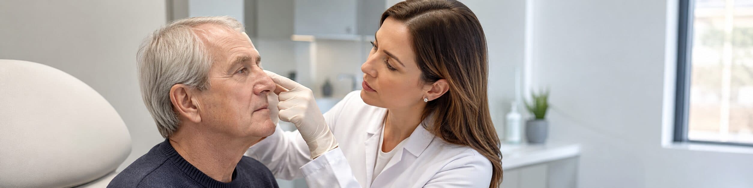
A clear guide to the most common skin cancer, its early signs, and usual management options.
Before you read: At Skinpaths, we distinguish between opinion and evidence. To write this content, we prioritized indexed medical sources and recommendations from scientific societies. You’ll find the complete bibliography at the end for your reference.
Important : This article is for informational purposes only and does not replace medical advice. If you have any questions, persistent symptoms, or your condition worsens, always consult a healthcare professional.
Summary in 60 seconds
- Basal cell carcinoma is a type of skin cancer that usually grows slowly.
- It appears more frequently in areas exposed to the sun, such as the face, nose, ears, scalp, neck, and arms.
- It may appear as a shiny «pearl», a wound that does not heal, a reddish plaque, a scar-like area, or a pigmented lesion.
- Although it rarely spreads to other organs, it can grow locally and damage nearby tissues if left untreated.
- Cumulative exposure to ultraviolet radiation is one of the most important factors.
- The diagnosis usually begins with clinical examination and can be confirmed with a biopsy.
- Treatment depends on the size, location, subtype, and risk of recurrence.
- Surgery is one of the most commonly used options; in selected cases, topical treatments, radiotherapy, photodynamic therapy, or systemic medications may be used.
- Consulting early allows for simpler treatments and better aesthetic and functional results.
- After a basal cell carcinoma, dermatological follow-up is important because new lesions may appear.
What is basal cell carcinoma?
Basal cell carcinoma is a skin cancer that originates in cells of the basal layer of the epidermis or in structures associated with hair follicles. It is considered the most common skin cancer in many populations, especially in people with fair skin and cumulative sun exposure. The American Academy of Dermatology guidelines describe it as the most common human skin cancer and a major proportion of non-melanoma skin cancers. Kim 2018
Its typical behavior is localized : it grows in the skin and nearby tissues. Metastasis is very rare, but this does not mean it is a «minor» lesion. A basal cell carcinoma located on the nose, eyelid, lips, ears, or scalp can compromise delicate structures if left untreated.
A wound that does not heal, bleeds, or reappears in the same spot deserves medical evaluation, even if it doesn’t hurt.
Why does it appear?
The most significant cause is cumulative damage from ultraviolet radiation , originating from the sun and, in some cases, tanning beds. Ultraviolet radiation can alter the DNA of skin cells; over time, some of these cells may proliferate abnormally.
The risk increases in people with fair skin, a history of sunburns, intense occupational or recreational sun exposure, advanced age, a personal history of skin cancer, immunosuppression, prior radiation therapy, or rare genetic syndromes. However, basal cell carcinoma can also occur in people with darker skin tones; in these individuals, some lesions may be pigmented and mistaken for moles or other spots. The Skin Cancer Foundation notes that in darker skin, a significant proportion of basal cell carcinomas may appear brown, black, or bluish.
Not all cases can be explained by «too much sun exposure.» Genetics, skin type, immune system, and history of radiation exposure also play a role. The available evidence supports a multifactorial view, not a single cause. Dika 2020
What does it look like and what symptoms does it cause?
Basal cell carcinoma can present in various ways. Some are subtle and are therefore mistaken for irritations, small wounds, pimples, scars, or spots.
It can be seen as:
- A small, shiny, translucent or «pearlescent» bump.
- A wound that bleeds, scabs over, partially heals, and reopens.
- A reddish or pinkish plaque that appears to be persistent irritation.
- A flat, firm, pale or yellowish area, similar to a scar.
- A lesion with raised edges and a more depressed center.
- A brown, black, or bluish stain, especially on more pigmented skin.
The American Cancer Society describes important warning signs as new or changing lesions, wounds that don’t heal, scaly patches, or areas that bleed, crust over, or recur. It also reminds us that not all skin cancers look the same.
Regarding symptoms, many lesions are painless . Some cause itching, tenderness, bleeding when touched, repeated crusting, or a feeling of a «wound that won’t heal.» The absence of pain does not rule out basal cell carcinoma.
What can you do today?
The safest approach is to systematically monitor your skin, avoiding self-medication, self-treatment, or attempts to «burn» or remove lesions at home. A helpful measure is to check areas exposed to the sun: face, ears, neck, scalp—especially if hair is sparse—chest, shoulders, arms, and backs of hands.
You can take photos in good light, without filters, and record the date. This helps to notice any real changes in size, color, bleeding, or shape. If a lesion doesn’t heal within a few weeks, bleeds for no clear reason, or reappears in the same spot, it’s advisable to seek medical evaluation from a dermatologist.
Sun protection is also part of sun care: seek shade, wear a hat, protective clothing, and use broad-spectrum sunscreen on exposed areas. These measures do not replace medical consultation when a suspected lesion is present, but they do reduce further ultraviolet damage.
What NOT to do?
It is not advisable to scratch, cut, squeeze, burn, or apply acids, home remedies, or «depigmenting» products to a suspicious lesion. These practices can irritate the area, delay diagnosis, and hinder clinical interpretation.
It is also unwise to assume that a lesion is not important because «it’s been the same for months» or because «it doesn’t hurt.» Many basal cell carcinomas grow slowly and can remain relatively stable for some time.
Avoid permanently covering a bleeding or crusted wound without seeking medical evaluation. Makeup, bandages, or creams can mask significant changes.
When should I consult a specialist?
Consult a dermatologist if any of these signs appear:
- A wound that does not heal after several weeks.
- A lesion that bleeds easily or forms repeated scabs.
- A shiny, pearly or translucent bump that grows.
- Persistent reddish plaque that does not improve.
- New or changing brown, black or blue spot.
- Injury to nose, eyelid, lips, ears, scalp, hands, genitals or scar area.
- Pain, rapid growth, deep ulceration, or repeated bleeding.
- Personal history of basal cell carcinoma, squamous cell carcinoma, or melanoma.
These signs don’t necessarily mean cancer, but they warrant evaluation. In cases of suspicious lesions, early consultation often allows for more conservative treatments.
What does a dermatologist usually do?
The dermatologist examines the lesion, asking about its duration, any changes, bleeding, history of skin cancer, sun exposure, immunosuppression, and previous treatments. They frequently use dermatoscopy , a non-invasive technique that allows them to view skin structures with magnification and special lighting.
If the lesion is suspicious, a biopsy may be indicated. A biopsy involves taking a small skin sample for histological confirmation. The technique depends on the size, depth, location, and clinical suspicion.
Once the diagnosis is confirmed, treatment is chosen based on risk. Factors that matter include location, size, borders, histological subtype, whether the lesion is new or recurrent, and the patient’s overall health. European and American guidelines agree that surgery is a mainstay of treatment, while non-surgical options are reserved for specific contexts, particularly low-risk superficial lesions or patients for whom surgery is not ideal. (Peris 2023 , Kim 2018)
Among the options that the specialist may consider are:
- Surgical excision: removing the lesion with a margin of healthy skin.
- Mohs micrographic surgery: a technique that analyzes the edges during the procedure; it is usually preferred in high-risk areas, recurrent lesions, or areas where preserving tissue is important.
- Curettage and electrodesiccation: an option for some small, low-risk basal cell carcinomas.
- Topical treatments: such as imiquimod or 5-fluorouracil, in selected superficial cases.
- Photodynamic therapy: an option in some superficial carcinomas.
- Radiotherapy: an alternative in selected patients or when surgery is not appropriate.
- Systemic therapies: reserved for advanced, inoperable, locally extensive or very rare cases with dissemination.
The American Cancer Society summarizes that the options depend on the size, location, general health, and patient preferences; it also notes that Mohs surgery can be especially useful for higher-risk tumors or those in areas such as the nose, eyes, ears, forehead, scalp, fingers, or genitals.
In darker skin tones, basal cell carcinoma can be pigmented and mistaken for a mole; change or persistence are important signs.
Frequently Asked Questions
Is basal cell carcinoma cancer?
- Yes. It is a type of skin cancer. However, it is usually less aggressive than melanoma and rarely metastasizes. Even so, it requires diagnosis and treatment because it can invade nearby tissues.
Can it just disappear?
- It’s not advisable to wait for it to disappear. Some lesions may seem to improve because they heal partially or crust over less, but then they reappear. This intermittent progression is a typical warning sign.
Hurts?
- It often doesn’t hurt. It may bleed, itch, crust over, or open with minimal trauma. The absence of pain does not mean it is benign.
Is it the same as melanoma?
- No. Basal cell carcinoma and melanoma are different skin cancers. Melanoma usually has a higher risk of spreading; basal cell carcinoma is usually localized, but can cause significant damage if it progresses.
Is surgery always necessary?
- Not always, but surgery is one of the most common options and offers the best control rates in many cases. For superficial and low-risk lesions, non-surgical treatments may be considered, at the doctor’s discretion.
Can I return after the treatment?
- Yes, it can recur, and new basal cell carcinomas can also appear in other areas of the skin. That’s why dermatological follow-up and sun protection are important.
What happens if it’s on the face?
- Lesions on the face, especially the nose, eyelids, lips, and ears, require careful evaluation. In these areas, the priority is usually to remove the tumor and preserve as much healthy tissue as possible, while also considering the functional and aesthetic outcome.
Bibliography and sources
- Kim JYS, Kozlow JH, Mittal B, Moyer J, Olencki T, Rodgers P, et al. Guidelines of care for the management of basal cell carcinoma. J Am Acad Dermatol . 2018;78(3):540-559. doi:10.1016/j.jaad.2017.10.006. Available at: https://pubmed.ncbi.nlm.nih.gov/29331385/
- Nasr I, McGrath EJ, Harwood CA, Botting J, Buckley P, Budny PG, et al. British Association of Dermatologists guidelines for the management of adults with basal cell carcinoma 2021. Br J Dermatol . 2021;185(5):899-920. doi:10.1111/bjd.20524. Available at: https://pubmed.ncbi.nlm.nih.gov/34050920/
- Peris K, Fargnoli MC, Kaufmann R, Arenberger P, Bastholt L, Seguin NB, et al. European consensus-based interdisciplinary guideline for diagnosis and treatment of basal cell carcinoma-update 2023. Eur J Cancer . 2023;192:113254. doi:10.1016/j.ejca.2023.113254. Available at: https://pubmed.ncbi.nlm.nih.gov/37604067/
- Peris K, Fargnoli MC, Garbe C, Kaufmann R, Bastholt L, Seguin NB, et al. Diagnosis and treatment of basal cell carcinoma: European consensus-based interdisciplinary guidelines. Eur J Cancer . 2019;118:10-34. doi:10.1016/j.ejca.2019.06.003. Available at: https://pubmed.ncbi.nlm.nih.gov/31288208/
- Queirolo P, Boutros A, Tanda ET, Spagnolo F, Quaglino P, et al. Guidelines for the diagnosis and treatment of basal cell carcinoma: a GRADE approach for evidence evaluation and recommendations by the Italian Association of Medical Oncology. ESMOOpen . 2023. Available at: https://pubmed.ncbi.nlm.nih.gov/37879235/
- Dika E, Scarfì F, Ferracin M, Broseghini E, Marcelli E, Bortolani B, et al. Basal Cell Carcinoma: A Comprehensive Review. Int J Mol Sci . 2020;21(15):5572. doi:10.3390/ijms21155572. Available at: https://pubmed.ncbi.nlm.nih.gov/32759706/
- van Loo E, Mosterd K, Krekels GAM, Roozeboom MH, Ostertag JU, Dirksen CD, et al. Surgical excision versus Mohs’ micrographic surgery for basal cell carcinoma of the face: a randomized clinical trial with 10 year follow-up. Eur J Cancer . 2014;50(17):3011-3020. doi:10.1016/j.ejca.2014.08.018. Available at: https://pubmed.ncbi.nlm.nih.gov/25262378/
- Chren MM, Linos E, Torres JS, Stuart SE, Parvataneni R, Boscardin WJ. Tumor recurrence five years after treatment of cutaneous basal cell carcinoma and squamous cell carcinoma. J Invest Dermatol . 2013;133(5):1188-1196. Available at: https://pmc.ncbi.nlm.nih.gov/articles/PMC3711403/
- American Academy of Dermatology. Basal cell carcinoma: guidelines. American Academy of Dermatology. Available at: https://www.aad.org/member/clinical-quality/guidelines/bcc
- American Cancer Society. Basal and Squamous Cell Skin Cancer Symptoms. American Cancer Society. Available at: https://www.cancer.org/cancer/types/basal-and-squamous-cell-skin-cancer/detection-diagnosis-staging/signs-and-symptoms.html
- American Cancer Society. Treating Basal Cell Carcinoma. American Cancer Society. Available at: https://www.cancer.org/cancer/types/basal-and-squamous-cell-skin-cancer/treating/basal-cell-carcinoma.html
- Skin Cancer Foundation. Basal Cell Carcinoma Overview. Skin Cancer Foundation. Available at: https://www.skincancer.org/skin-cancer-information/basal-cell-carcinoma/
Categories: For everyone · Skin cancer

Content reviewed by
Dr. Rodolfo Suárez
Médico Patólogo y Dermatólogo Master en Patología Cutánea Avanzada
- Published
- September 18, 2026
- Updated
- September 23, 2026
Medically reviewed by specialists
Our content is based on current scientific evidence and clinical protocols.