Sweet's syndrome

Directory of diseases

Sweet's syndrome

Created: 12/01/2026 Last updated: 10/01/2026 Important: This content is for informational purposes only and is intended for healthcare professionals. It does not replace clinical assessment or medical judgment, does not establish a doctor-patient relationship, and does not constitute a standard of care. Medicine is rapidly evolving: always verify sources and use this information only as additional input for decision-making.

General

  • Synonyms: Acute febrile neutrophilic dermatosis (DNFA), (acute febrile neutrophilic dermatosis).
  • Definición: inflammatory dermatosis characterized by abrupt start de painful erythematous-violaceous papules/nodules/plaques, often accompanied by fever y elevated inflammatory markers, with neutrophilic dermal infiltrate (typically without primary vasculitis). Sweet 1964, Cohen 2007
  • First description: Robert Douglas Sweet, 1964 (Br J Dermatol). Sweet 1964
  • Epidemiology (practical pearls):
    • More frequent in of middle-aged in the “classic/idiopathic” form. von den Driesch 1994, Cohen 2007
    • It can be associated with neoplasms (especially hematological diseases such as AML/MDS) and in a subgroup precedes or coincideswith the cancer diagnosis. Cohen 1988, Bourke 1997
    • Pregnancy: recognized association; modern reviews summarize presentation and approach in gestation. von den Driesch 1994, Glennon 2024
    • Codes (indicative):
      • ICD-10: is usually coded as L98.2 (other skin and subcutaneous tissue conditions).
      • ICD-11: It may vary by local mapping (search “Sweet syndrome / acute febrile neutrophilic dermatosis” in the institutional ICD-11 browser).
  • Diagnostic checklist (quick)
    • Clinical presentation: abrupt onset of painful lesions + fever/malaise. Cohen 2007
    • Labs: Complete blood count with differential + CRP/ESR. Fat 1995
    • Confirmation: punch biopsy. Guhl 2008
    • Criteria: apply set of criteria (Su/Liu and modern revisions when “classic” elements are missing). On 1986, Nofal 2017
    • Targeted screening: recent drugs (G-CSF/ATRA/others) and search for malignancy if red flags are present. Thompson 2007, Cohen 1988
  • Three main clinical contexts:
    1. Classic/idiopathic (sometimes post-infectious or associated with IBD/pregnancy). Cohen 2007 , von den Driesch 1994

    2. Associated with malignancy (especially hematological; may be accompanied by cytopenias). Cohen 1988 , Bourke 1997

    3. Drug-induced (classically G-CSF* ; also ATRA** and others). Thompson 2007

  • Pathophysiology: hypersensitivity/cytokine reaction with neutrophil recruitment; response to blockade of IL-1 It supports an inflammasome-IL-1 axis in refractory cases. Cohen 2007, Kluger 2011

*G-CSF: graulocite colony-stimulating factor, ** ATRA: all-trans retinoic acid


  • Elementary injury
  • Typical areas
  • prototypical picture
    • Abrupt onset + fever/malaise ± arthralgia, with neutrophilia and/or elevated ESR/CRP. Cohen 2007, Fat 1995
  • Extracutaneous findings (key to oculoplastics/internal medicine)
    • Eyepiece: conjunctivitis, episcleritis/scleritis, ulcerative peripheral keratitis, iritis/uveitis; also orbital/periorbital inflammation (may mimic orbital cellulitis). Gottlieb 2008
    • Neurological: "neuro-Sweet"(aseptic meningoencephalitis/CNS manifestations) with proposed criteria. Hisanaga 2005
  • Evolution
  • Important atypical forms
    • Subcutaneous (neutrophilic panniculitis): predominance in subcutaneous cellular tissue; relevant relationship with hematological malignancy; may be confused with erythema nodosum. Guhl 2008
    • Histiocytoid: infiltrate of cells with histiocytic appearance but MPO+ (immature granulocytes); relatively greater association with MDS in comparative series. Requena 2005, Ghoufi 2016
    • Variants that mimic necrotizing fasciitis: “neutrophilic dermatosis variants resembling necrotizing fasciitis” → risk of unnecessary debridement if mistaken for infection. sanchez 2019
  • Cellulitis/erysipelas: similar pain and fever, but there is usually an infectious focus/portal of entry and a response to antibiotics; in Sweet's syndrome, the biopsy shows a sterile neutrophilic infiltrate and a response to steroids. Cohen 2007

  • Leukocytoclastic vasculitis: palpable purpura, necrosis; histology with primary vasculitis (secondary vascular changes may be present in Sweet's disease). von den Driesch 1994

  • Pyoderma gangrenosum: ulceration with an undermined border and pathergy; it may coexist within the spectrum of neutrophilic dermatoses. Cohen 2007

  • Erythema nodosum: painful pretibial nodules; septal histology (subcutaneous sweetness = neutrophilic panniculitis). Guhl 2008

  • Cutaneous leukemia/tumor infiltration: suspect if atypical lesions + cytopenias/blasts; in histiocytoid, use IHC (MPO/CD15) and hematological correlation. Requena 2005 , Cohen 1988

  • Histological pattern
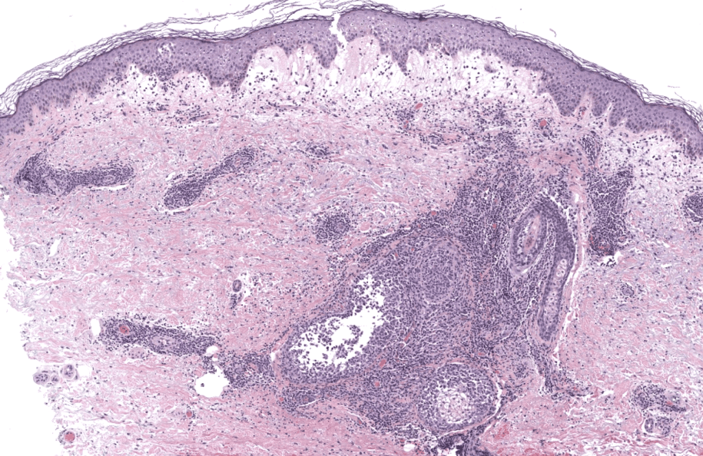
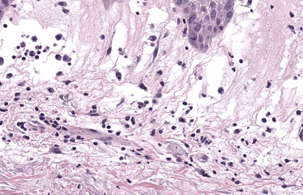
    • Neutrophilic dermatitis: dense neutrophilic infiltrate in the dermis (frequent papillary edema) with leukocytoclasia; typically without primary vasculitis. von den Driesch 1994, Cohen 2007
  • Key findings
    • Classic: neutrophil-rich inflammatory infiltrate in the dermis; edema. Cohen 2007
    • Histiocytoid: “histiocytoid” cells but intensely positive MPO (immature granulocytes). Requena 2005
    • Subcutaneous: neutrophilic panniculitis (sometimes predominantly lobular). Guhl 2008
  • Useful IHC/stains (depending on differential)
    • To rule out infection when there is clinical doubt: Gram, PAS/GMS, Ziehl-Neelsen (and cultivation if applicable). Cohen 2007
    • If histiocytoid/leukemia cutis is suspected: MPO, CD15, correlation with hematology; some cases require additional studies. Requena 2005, Ghoufi 2016
  • molecular tests
    • They are not routine for "classic" Sweet; they are reserved for suspected hematological neoplasia or alternative diagnoses. Cohen 1988, Ghoufi 2016


Sweet's syndrome: This is a neutrophilic dermatosis where there is usually papillary dermal edema associated with a dense inflammatory infiltrate of neutrophilic predominance with a tendency to coalesce around vascular plexuses and glandular structures


Sweet's syndrome: papillary dermal edema with neutrophilic infiltrate
  • Infection (bacterial, fungal, mycobacterial): neutrophils + microorganisms/positive stains/cultures. Cohen 2007

  • Primary leukocytoclastic vasculitis: fibrinoid damage and neutrophils around vessels as the primary event. von den Driesch 1994

  • Cutaneous leukemia: blastic infiltrate; in histiocytoid leukemia, MPO+ may be confusing—correlate with clinical presentation/blood count. Requena 2005

  • Basic initial (most patients):
    • Complete blood count with differential (look for neutrophilia or cytopenias). Fat 1995, Cohen 1988
    • PCR/ESR (inflammatory activity). Fat 1995
    • Liver/kidney function (before colchicine/dapsone/cyclosporine if contemplated). Cohen 2002
  • If malignancy or histiocytoid/subcutaneous variant is suspected:
    • Peripheral smear ± hematological assessment; in histiocytoid, hematological evaluation and follow-up of counts are recommended. Cohen 1988, Ghoufi 2016
  • If there is extracutaneous data:
    • Urgent ophthalmology If you experience eye pain, photophobia, or decreased vision. Gottlieb 2008
    • Neuroimaging/CSF if meningoencephalitic syndrome compatible with neuro-Sweet (according to criteria). Hisanaga 2005
  • Biopsy
    • Recommended type
      • Punch 4–6 mm deep (include reticular dermis; add subcutaneous cellular tissue (if subcutaneous form is suspected). Guhl 2008, Cohen 2007
    • Key considerations
      • Choosing early injury and active (not just ulcerated/necrotic area). Cohen 2007
      • If the differential includes infection: take a sample for culture separately (sterile container) in addition to the piece in formalin. Cohen 2007
    • Contraindications
      • Not specific; apply clinical judgment (anticoagulation, anatomical site, etc.).
    • Fixing/Shipping
      • Histology: 10% formalin
  • front line
    • Systemic corticosteroid (e.g., prednisone/prednisolone tapering): gold standard and usually gives a rapid response. Cohen 2002, Cohen 2007
    • Topical or intralesional (adjuvant or limited cases). Cohen 2002
  • Alternatives (when steroids are risky or as a cost-saving measure)
    • Colchicine o potassium iodideOral options with reported rapid response. Cohen 2002,
    • Other options with clinical experience: indomethacin, dapsone, cyclosporine, clofazimine (select by comorbidities). Cohen 2002
    • Refractory: blocking IL-1 (anakinra) with marked responses in reports, useful as a proof of concept. Kluger 2011
  • Care/monitoring (high performance)
    • Before dapsoneG6PD, hemolysis/methemoglobinemia, and serial blood counts (if used). (Practical guide within therapeutic review). Cohen 2002
    • Colchicine: Monitor for cytopenias, GI toxicity, and adjustments for renal/hepatic function and interactions. Cohen 2002
    • Potassium iodide: Monitor for GI intolerance, iodine levels, and thyroid function in prolonged/high-risk treatments. Horio 1980, Cohen 2002
  • Contraindications (practical)
    • Steroids: caution in uncontrolled infection; prioritize ruling out sepsis/infectious necrosis if the clinical picture suggests it. Cohen 2007, sanchez 2019
    • Pregnancy: individualize (specific review during pregnancy). Glennon 2024
  • Suspicion of underlying neoplasia : older age, male sex, cytopenias , severe/atypical lesions, histiocytoid or subcutaneous → hematological evaluation and follow-up. Cohen 1988 , Bourke 1997 , Ghoufi 2016 , Guhl 2008

  • Painful red eye / photophobia / decreased vision : risk of keratitis/uveitis/scleritis → ophthalmological emergency. Gottlieb 2008

  • A clinical picture that “resembles necrotizing fasciitis” (disproportionate pain, purplish plaques, fever, antibiotic resistance): consider neutrophilic variant type NF; avoid repeated debridement without confirming infection. Sanchez 2019

  • Neurological symptoms (severe headache, confusion, neck stiffness): consider NeuroSweet and apply diagnostic criteria. Hisanaga 2005

  • Painful , erythematous-violaceous , “pseudovesicular” plaques with a rapid response to steroids are highly suggestive (after excluding infection). von den Driesch 1994 , Cohen 2002

  • Histiocytoid staining may appear as "something else" under H&E; MPO+ provides guidance. Requena 2005

  • Sweet RD. AN ACUTE FEBRILE NEUTROPHILIC DERMATOSIS. Br J Dermatol. 1964; 76: 349-356. DOI:10.1111/j.1365-2133.1964.tb14541.x. PMID: 14201182.
    Brief summary: Foundational publication that describes the original clinical picture of acute febrile neutrophilic dermatosis, establishing the clinical phenotype and the entity that would later bear the eponym.
    Sweet 1964
  • Your WP, Liu HN. Diagnostic criteria for Sweet's syndrome. Skin. 1986;37(3):167–174. DOI: Not available in PubMed. PMID: 3514153.
    Brief summary: It proposes major and minor criteria for a "defined" diagnosis, having a lasting influence on clinical-histological diagnostic standardization.
    On 1986

  • von den Driesch P. Sweet's syndrome (acute febrile neutrophilic dermatosis). J Am Acad Dermatol.1994;31(4):535–556; quiz 557–560. DOI: 10.1016/S0190-9622(94)70215-2. PMID: 8089280.
    Brief summary: Classic review that systematizes clinical presentation (including pseudovesicular appearance), associations (including pregnancy) and diagnostic-therapeutic approach, with a broad view of extracutaneous manifestations.
    von den Driesch 1994

  • Fett DL, Gibson LE, Su WP. Sweet's syndrome: systemic signs and symptoms and associated disorders. Mayo Clin Proc. 1995;70(3):234–240. DOI: 10.4065/70.3.234. PMID: 7861810.
    Brief summary: Institutional series (Mayo Clinic) that characterizes systemic signs, associated comorbidities and response to treatment, useful for estimating the “systemic burden” beyond the skin.
    Fat 1995

  • Bourke JF, Keohane S, Long CC, et al. Sweet's syndrome and malignancy in the UK Br J Dermatol.1997;137(4):609–613. DOI: 10.1111/j.1365-2133.1997.tb03796.x. PMID: 9390341.
    Brief summary: It analyzes the Sweet-malignancy relationship in the United Kingdom, providing clinical data applicable to cancer screening decisions and risk profiling.
    Bourke 1997

  • Cohen PR, Talpaz M, Kurzrock R. Malignancy-associated Sweet's syndrome: review of the world literature. J Clin Oncol. 1988;6(12):1887–1897. DOI: 10.1200/JCO.1988.6.12.1887. PMID: 3058878.
    Brief summary: Seminal review that consolidates the association with cancer (particularly hematological), describes clinical clues (e.g., cytopenias) and highlights that Sweet may precede or coincide with the oncological diagnosis.
    Cohen 1988

  • Cohen PR, Kurzrock R. Sweet's syndrome: a review of current treatment options. Am J Clin Dermatol.2002;3(2):117–131. DOI: 10.2165 / 00128071-200203020-00005. PMID: 11893223.
    Brief summary: Practical therapeutic review: corticosteroids as standard, and alternatives (colchicine, potassium iodide, indomethacin, dapsone, cyclosporine), with clinical guidance for scenarios where steroid is limited.
    Cohen 2002

  • Cohen PR. Sweet's syndrome—a comprehensive review of an acute febrile neutrophilic dermatosis. Orphanet J Rare Dis. 2007; 2: 34. DOI: 10.1186/1750-1172-2-34. PMID: 17655751. PMCID: PMC1963326.
    Brief summary: Comprehensive review (with full text in PMC) that integrates subtypes (classic, malignancy, drugs), clinical presentation, pathology, extracutaneous manifestations, and recurrences; excellent as a framework reference.
    Cohen 2007

  • Thompson DF, Montarella KE. Drug-induced Sweet's syndrome. Ann Pharmacother. 2007;41(5):802–811. DOI:10.1345/aph.1H563. PMID: 17426076.
    Brief summary: Systematic review focused on pharmacovigilance; discusses strength of association by drug (highlighting G-CSF/ATRA) and proposes a causality approach useful in practice.
    Thompson 2007

  • Nofal A, Abdelmaksoud A, Amer H, et al. Sweet's syndrome: diagnostic criteria revisited. J Dtsch Dermatol Ges. 2017;15(11):1081–1088. DOI: 10.1111/ddg.13350. PMID: 28981179.
    Brief summary: Re-evaluate diagnostic criteria in the face of incomplete or atypical presentations, providing a modern framework for "non-textbook" cases.
    Nofal 2017

  • Gottlieb CC, Mishra A, Belliveau D, et al. Ocular involvement in acute febrile neutrophilic dermatosis (Sweet syndrome): new cases and review of the literature. Surv Ophthalmol. 2008;53(3):219–226. DOI:10.1016/j.survophthal.2008.02.006. PMID: 18501268.
    Brief summary: Key review to recognize the broad ocular spectrum (from conjunctivitis to uveitis/scleritis and orbital involvement) and prioritize urgent referral when appropriate.
    Gottlieb 2008

  • Hisanaga K, Iwasaki Y, Itoyama Y; Neuro-Sweet Disease Study Group. Neuro-Sweet disease: clinical manifestations and criteria for diagnosis. neurology. 2005;64(10):1756–1761. DOI:10.1212/01.WNL.0000161848.34159.B5. PMID: 15911805.
    Brief summary: It defines neuro-Sweet as an entity with CNS involvement and proposes diagnostic criteria, facilitating differentiation from other neuroinflammatory syndromes.
    Hisanaga 2005

  • Sanchez IM, Lowenstein S, Johnson KA, et al. Clinical Features of Neutrophilic Dermatoses Variants Resembling Necrotizing Fasciitis. JAMA Dermatol. 2019;155(1):79–84. DOI: 10.1001/jamadermatol.2018.3890. PMID: 30383110. PMCID: PMC6439570.
    Brief summary: Multicenter study characterizing neutrophilic variants that mimic necrotizing fasciitis; essential to prevent aggressive surgery when the process is sterile inflammation.
    sanchez 2019

  • Guhl G, García-Díez A. Subcutaneous sweet syndrome. Dermatol Clin. 2008;26(4):541–551, viii–ix. DOI:10.1016/j.det.2008.06.003. PMID: 18793988.
    Brief summary: Review of subcutaneous Sweet's disease within the spectrum of neutrophilic panniculitis, with emphasis on differentials (erythema nodosum) and association with hematological malignancy.
    Guhl 2008

  • Requena L, Kutzner H, Palmedo G, et al. Histiocytoid Sweet syndrome: a dermal infiltration of immature neutrophilic granulocytes. Arch Dermatol. 2005;141(7):834–842. DOI: 10.1001/archderm.141.7.834. PMID:16027297.
    Brief summary: Key series that defines the histiocytoid subtype and demonstrates by IHC (MPO, CD15 and others) that “histiocytoid” cells correspond to immature granulocytes, with diagnostic and oncohematological implications.
    Requena 2005

  • Ghoufi L, Ortonne N, Ingen-Housz-Oro S, et al. Histiocytoid Sweet Syndrome… A Comparative Series of 62 Patients. Medicine (Baltimore). 2016, 95 (15): e3033. DOI: 10.1097/MD.0000000000003033. PMID: 27082547. PMCID: PMC4839791.
    Brief summary: Extensive comparison of histiocytoid vs. classic histiocytoids shows a significant association with MDS and recommends hematological evaluation and follow-up of counts, providing practical evidence for “red flags”.
    Ghoufi 2016

  • Horio T, Imamura S, Danno K, et al. Treatment of acute febrile neutrophilic dermatosis (Sweet's Syndrome) with potassium iodide. Dermatological. 1980;160(5):341–347. DOI: 10.1159/000250516. PMID: 7364144.
    Brief summary: It reports the usefulness of potassium iodide as a therapeutic alternative, relevant when steroids are contraindicated or a rapid response is sought with another route.
    Horio 1980

  • Kluger N, Gil-Bistes D, Guillot B, Bessis D. Efficacy of… anakinra… in a case of refractory Sweet's syndrome.Dermatology. 2011;222(2):123–127. DOI: 10.1159/000326112. PMID: 21464561.
    Brief summary: Refractory case with marked response to anakinra; supports the role of IL-1/inflammasome and paves the way for targeted therapies in difficult neutrophilic dermatoses.
    Kluger 2011

  • Glennon CM, Tan AJ, Prabhu M, Kroshinsky D. Sweet syndrome in pregnancy: A narrative review. International Journal of Gynaecology and Obstetrics. 2024;166(2):538–550. DOI: 10.1002/ijgo.15713. PMID: 38881204.
    Brief summary: Review focused on pregnancy: presentation, relevant differential diagnoses and therapeutic considerations/maternal-fetal safety, useful for multidisciplinary management.
    Glennon 2024

We know skin / We understand skin
© 2026 Skinpaths. All rights reserved.