
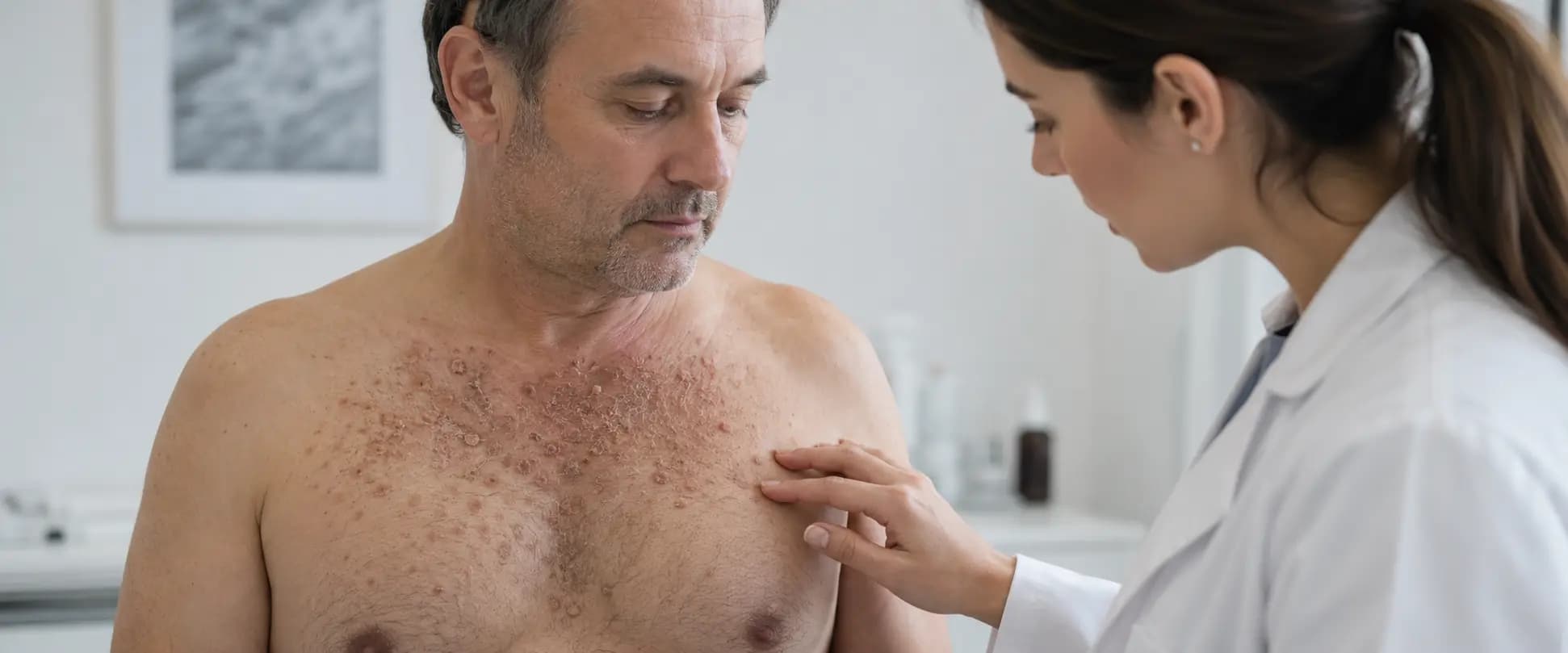
A genetic and chronic skin disease that can be controlled through outbreak prevention, infection surveillance, and individualized treatment.
Before you read: At Skinpaths, we distinguish between opinion and evidence. To write this content, we prioritized indexed medical sources and recommendations from scientific societies. You’ll find the complete bibliography at the end for your reference.
Important: This article is for informational purposes only and does not replace medical advice. If you have any questions, persistent symptoms, or your condition worsens, consult a healthcare professional.
Summary in 60 seconds
- Darier disease is a rare genetic disorder that primarily affects the skin, nails, and sometimes the mouth.
- It is related to variants of the ATP2A2 gene, which is responsible for producing a protein that regulates calcium within cells.
- It is not contagious, it is not due to an allergy, and it does not appear due to a lack of hygiene.
- It usually begins during late childhood, adolescence, or early adulthood.
- It produces small, rough or crusty bumps, especially on the chest, back, scalp, neck, and skin folds.
- Heat, sweat, humidity, friction, and ultraviolet radiation can all contribute to outbreaks.
- The diagnosis is usually clinical, although a biopsy or genetic test may be necessary.
- Currently there is no treatment that produces a permanent remission, but there are options to control the lesions and reduce relapses.
- A sudden worsening, severe pain, fever, blisters, or extensive erosions requires prompt medical evaluation.
What is Darier’s disease?
Darier’s disease, also called Darier-White disease or follicular dyskeratosis , is a genodermatosis: a skin disease related to a genetic alteration.
It affects how epidermal cells adhere to each other and complete their maturation process. As a result, the skin barrier becomes more fragile, and thickened, rough, inflamed, or crusty areas may appear.
It is a rare disease. Prevalence estimates vary among populations, although it is generally considered to affect a few people per 100,000 inhabitants. It usually follows a chronic course, with relatively quiet periods and flare-ups of varying intensity.
It is not transmitted by touching the lesions, sharing clothing, or living with an affected person. It is also not caused by an allergy, an initial infection, or poor hygiene. The former name “follicular keratosis” may be inaccurate, as the lesions do not necessarily originate in the hair follicles. (Has 2026, DermNet 2023 )
Why does it appear?
The primary cause is a pathogenic variant in the ATP2A2 gene. This gene contains the instructions for producing SERCA2 , a pump that regulates the movement of calcium within cells.
Calcium is important for skin cells to bind together and mature properly. When SERCA2 is not functioning correctly, cohesion between keratinocytes decreases, and their keratinization is impaired. This helps explain the fragility, thickening, and inflammation characteristic of the disease.
How is it inherited?
The generalized form usually follows an autosomal dominant pattern. When one parent carries a germline variant of ATP2A2, each pregnancy has a 50% chance of inheriting it. This figure refers to genetic transmission; it does not allow for predicting the severity of the disease.
Two members of the same family can present very different manifestations. One person may have subtle lesions while another experiences extensive outbreaks, even while sharing the same genetic variant.
The absence of a family history does not rule out the diagnosis. The abnormality may have appeared for the first time in that person, gone unnoticed in relatives with very mild symptoms, or be present only in a portion of the cells, as occurs in some segmental forms.
Triggers are not the cause
Heat, sweat, or sun exposure do not cause Darier’s disease. However, they can trigger or intensify lesions in genetically susceptible skin. The most commonly described triggers are:
- heat and humidity;
- profuse sweating;
- ultraviolet exposure;
- Tight clothing, chafing or scratching;
- skin infections;
- hormonal changes;
- physical or emotional stress;
- some medications.
Lithium is one of the drugs most clearly associated with exacerbations. It should not be discontinued abruptly: any possible relationship between a medication and flares should be reviewed with the prescribing professional. Sakuntabhai 1999, Has 2026 .
Triggers do not cause the disease: they can trigger outbreaks in genetically susceptible skin.
What does it look like and what symptoms does it cause?
The most representative lesion is the papule , that is, a small, firm, palpable bump. It usually has a rough, scaly, waxy, or crusty surface. Its color can be reddish, yellowish, brown, or close to the natural skin tone.
The papules may appear singly or merge to form thicker, irregular, wart-like plaques. Their appearance varies depending on skin pigmentation, location, the presence of inflammation, and the existence of a secondary infection.
Most frequent areas
The lesions tend to be concentrated in areas with higher oil production or a tendency towards moisture:
- scalp and hairline;
- forehead, temples and region around the ears;
- neck;
- central area of the chest;
- upper back;
- armpits;
- English;
- fold located below the breasts.
Maceration, fissures, erosions, and exudate may appear in skin folds. These areas are especially vulnerable to overgrowth of bacteria, yeast, and viruses.
Possible symptoms
The severity varies greatly. Some people have minor, virtually asymptomatic lesions; others may experience:
- itching;
- burning sensation;
- pain or sensitivity;
- cracks;
- moisture or exudate;
- unpleasant smell;
- recurrent infections.
The odor is usually related to moisture, maceration, and microbial growth on a compromised skin barrier. It should not be automatically interpreted as a lack of hygiene.
Changes in the nails, hands, and mouth
The nails provide important diagnostic clues. The following may be observed:
- red or white longitudinal lines;
- fragility of the nail plate;
- small “V” shaped notches on the free edge.
Small pits or hardened spots may also appear on the palms and soles, as well as flat, wart-like papules on the backs of the hands.
In some people, small whitish bumps form on the palate, gums, tongue, or inner cheeks. When numerous, the mucous membrane can take on a cobblestone-like texture.
A sudden increase in pain, with or without extensive blistering, erosion, or crusting, should raise suspicion of infection. Superinfection with herpes simplex is a particularly important complication. Ettinger 2025 , BAD 2020 .
What can you do today?
The following measures do not modify the genetic alteration, but they can decrease exposure to triggers and protect the skin barrier.
1. Reduce heat and sweating
On hot days, try to stay in well-ventilated areas, take breaks to cool down, and change out of wet clothes after sweating. Exercise is not prohibited, but you may need to adjust its intensity, timing, and environment.
2. Choose breathable clothing
Loose, soft clothing reduces chafing and occlusion. Stiff, tight, or poorly ventilated fabrics can increase moisture in skin folds.
3. Practice consistent sun protection
Seek shade, wear protective clothing, and apply a broad-spectrum sunscreen with SPF 30 or higher to exposed areas. Choose well-tolerated formulas, especially if your skin is inflamed.
4. Clean without harming
Use lukewarm water and a mild cleanser without strong perfumes. Avoid brushes, rough sponges, and vigorous rubbing. After bathing, pat your skin dry, paying attention to skin folds.
5. Protect the skin barrier
A simple, fragrance-free emollient can reduce dryness, irritation, and some flaking. Products containing urea, lactic acid, or other keratolytic agents can be helpful on thickened areas, but they can also cause stinging and are not suitable for all locations.
6. Observe the evolution
Recording photographs, affected areas, ambient temperature, perspiration, menstrual cycle, infections, and products used can help identify personal patterns. It’s advisable to check your skin regularly for new pain, erosions, pus, or rapid changes.
7. Follow a medical plan for outbreaks
If a diagnosis has already been made, ask for clear instructions on what treatment to use at the onset of an outbreak and what signs require a consultation. Antiseptic washes may be helpful in certain patients, but their use should be individualized according to medical criteria to avoid irritation or sensitization. Has 2026, BAD 2020 .
The realistic goal of treatment is to reduce injury, discomfort, and infection, not to promise a definitive cure.
What NOT to do?
- Do not scrape, sand, or pick at scabs. Trauma can increase inflammation, break down the skin barrier, and make infections more likely.
- Do not use the sun to «dry out» the lesions. Ultraviolet radiation is a common trigger.
- Do not apply retinoids, strong acids, or potent corticosteroids on your own. The choice depends on the affected area, the degree of inflammation, and your personal medical history.
- Do not use topical antibiotics for prolonged or repeated periods without medical advice. This can promote bacterial resistance and contact dermatitis.
- Do not cover a wet or eroded area occlusively , unless a professional has indicated a specific dressing.
- Do not discontinue lithium or any other prescribed medication on your own. Any change should be coordinated with the doctor treating the condition for which it was prescribed.
- Do not attribute every worsening to a “normal flare-up.” A sudden change to increased pain, blisters, erosions, or fever may indicate an infection.
The most recent consensus recommendations emphasize that care should be tailored to the predominant type of lesion: keratosis, inflammation, moisture, infection, or a combination of these factors. (Has 2026 )
When should I consult?
Seek medical attention the same day or go to the emergency room if the following occurs:
- rapid worsening and much more painful than usual , even if no blisters are visible;
- clustered vesicles, small blisters, extensive erosions or lesions with a “squashed” appearance;
- fever, chills, general malaise, or swollen lymph nodes;
- redness that spreads, intense local heat, swelling or pus;
- abundant exudate or new odor accompanied by pain;
- extensive skin involvement, weakness or difficulty maintaining adequate hydration;
- eye pain, red eye, intense sensitivity to light, or changes in vision;
- a lesion that grows persistently, ulcerates, bleeds, or does not heal.
The combination of severe pain, erosions, and rapid deterioration may indicate disseminated herpes simplex infection. This complication requires urgent evaluation and antiviral treatment when confirmed or when there is sufficient clinical suspicion.
Schedule a dermatology appointment if:
- The injuries are new and there is no diagnosis;
- Outbreaks are frequent or increasingly widespread;
- The itching, pain, or odor affect sleep, work, studies, or personal relationships;
- Standard treatments no longer control the symptoms;
- repeated infections appear;
- The illness affects mood or promotes social isolation;
- you wish to plan a pregnancy;
- You need to know the risk to family members or children.
What does a dermatologist usually do?
Medical history and examination
The dermatologist examines the distribution and morphology of the lesions, as well as the nails, palms, soles, and oral mucosa. They also usually ask about:
- age of onset;
- family history;
- relationship with heat, sun, sweat or friction;
- previous infections;
- medications, especially lithium;
- impact on sleep, work, relationships and mood.
Dermatoscopy can provide additional information about the keratin and architecture of the lesions, although it is not always necessary.
Skin biopsy
When the appearance is atypical or it is necessary to differentiate the disease from other disorders, a punch biopsy may be performed: a small cylinder of skin is removed under local anesthesia. Microscopic examination usually shows loss of adhesion between cells—acantholysis—and abnormal maturation of keratinocytes—dyskeratosis. These findings must be interpreted in conjunction with the clinical examination. Diagnoses that may be considered include Hailey-Hailey disease, Grover’s disease, seborrheic dermatitis, and certain verrucous or epidermal lesions.
Infection screening
If there is oozing, crusting, a new odor, or pain, samples can be taken for bacterial or fungal culture. If herpes simplex is suspected, the doctor may order a PCR test or start antiviral treatment without waiting for the result if the symptoms warrant it.
Genetic study
ATP2A2 testing can support diagnosis and facilitate family counseling. It is particularly useful in atypical, segmental, early-onset presentations, or those without a clear family history. A negative test does not always rule out the disease, as some variants may not be detected by all available methods. In segmental forms, it is sometimes necessary to test both blood and affected skin.
Individualized treatment
In mild or localized cases, the dermatologist may use a combination of:
- emollients;
- keratolytics;
- selected antiseptics;
- topical corticosteroids during short periods of inflammation;
- calcineurin inhibitors in sensitive areas or folds;
- carefully introduced topical retinoids.
When an infection is present, treatment targets the responsible microorganism with antibacterial, antifungal, or antiviral drugs. Not all exudate requires antibiotics. In extensive, persistent, or severely debilitating cases, oral retinoids , such as acitretin or isotretinoin, remain the most clinically established systemic treatment options. They can reduce the thickening and number of lesions, but they do not correct the underlying genetic alteration, and relapses after discontinuation are common. These medications require monitoring, laboratory tests, and a thorough evaluation of their potential adverse effects. They are highly teratogenic, therefore requiring prescription and follow-up by a specialist physician and strict protocols for preventing pregnancy.
Localized, refractory lesions may be evaluated for procedures such as ablative laser, dermabrasion, or surgery. The choice depends on the location, type of lesion, and risk of scarring. Therapies targeting JAK, IL-17, IL-23, IL-4/IL-13, or MEK pathways, as well as potential gene therapy strategies, are still under investigation or primarily based on small case series. They should not be presented as routine treatments or guaranteed solutions. The ERN-Skin international consensus (2026) supports a stepped but flexible approach: preventing triggers and superinfections, treating the predominant component in each patient, and reserving systemic or procedural therapies for cases that require them. ( Has 2026 , Hanna 2022 )
A sudden change towards increased pain, erosions, or fever warrants prompt medical evaluation.
Frequently Asked Questions
Is Darier’s disease contagious?
- No. It is a genetic disease and cannot be transmitted through physical contact, sexual relations, swimming pools, clothing, towels, or cohabitation.
Is it an autoimmune disease or an allergy?
- It is not classified as an autoimmune or allergic disease. Its origin lies in an alteration of ATP2A2. Inflammation and infections can complicate the lesions, but they are not the genetic cause.
Will all children of an affected person develop the disease?
- No. In the autosomal dominant form, each pregnancy has a 50% chance of inheriting the variant. Furthermore, the severity cannot be predicted by the severity observed in the other parent. Genetic counseling allows for a more precise study of each family situation. (Has 2026 )
Is there a cure?
- Currently, there is no therapy that produces permanent remission. However, many people achieve long periods of control through trigger prevention, skin care, and treatments tailored to the extent and type of lesions.
Can retinoids be used during pregnancy?
- Oral retinoids are highly teratogenic and are contraindicated during pregnancy. Topical retinoids should also be avoided except in exceptional, specialized cases. Planning should be coordinated with dermatology and obstetrics, following local protocols for each medication.
Is Darier’s disease the same as Darier’s sign?
- No. Darier’s sign describes the appearance of redness, itching, and swelling when certain mastocytosis lesions are rubbed. It shares the eponym, but does not correspond to the genetic disease Darier. Surjushe 2007 .
Can it affect anything other than the skin?
- It can affect the nails and oral mucosa. Some people experience eye discomfort, dental problems, or significant impact on their emotional health. Neuropsychiatric associations have also been described, but this does not mean that everyone will develop these problems. Follow-up should be tailored to each patient’s specific symptoms.
Bibliography and sources
- Has C, Ruffo di Calabria V, Caproni M, Didona B, Fischer J, Gostynski A, et al. Consensus Recommendations for Management of Darier Disease: A Practical Approach . Dermatol Ther (Heidelb). 2026 Jul 15. doi: 10.1007/s13555-026-01838-3.
- Ettinger M, Kimeswenger S, Deli I, Traxler J, Altrichter S, Noack P, et al. Darier disease: Current insights and challenges in pathogenesis and management . J Eur Acad Dermatol Venereol. 2025;39(5):942-951. doi: 10.1111/jdv.20448.
- Hanna N, Lam M, Fleming P, Lynde CW. Therapeutic Options for the Treatment of Darier’s Disease: A Comprehensive Review of the Literature . J Cutan Med Surg. 2022;26(3):280-290. doi: 10.1177/12034754211058405.
- Haber RN, Dib NG. Management of Darier disease: A review of the literature and update . Indian J Dermatol Venereol Leprol. 2021;87(1):14-21. doi: 10.25259/IJDVL_963_19.
- Sakuntabhai A, Ruiz-Perez V, Carter S, Jacobsen N, Burge S, Monk S, et al. Mutations in ATP2A2, encoding a Ca2+ pump, cause Darier disease . Nat Genet. 1999;21(3):271-277. doi:10.1038/6784.
- British Association of Dermatologists. Darier disease [Internet] . London: British Association of Dermatologists; 2020 Dec [citado 18 jul 2026] .
- Primary Care Dermatology Society. Darier’s disease [Internet] . Primary Care Dermatology Society; updated May 28, 2022 [citado 18 jul 2026] .
- DermNet New Zealand. Darier disease [Internet] . Hamilton: DermNet New Zealand; updated Nov 30, 2023 [citado 18 jul 2026] .
- Orphanet. Darier disease. ORPHA:218 [Internet] . Paris: INSERM [citado 18 jul 2026] .
- MedlinePlus Genetics. Darier disease [Internet] . Bethesda (MD): National Library of Medicine; updated Jan 12, 2024 [citado 18 jul 2026] .
- Surjushe A, Jindal S, Gote P, Saple DG. Darier’s sign . Indian J Dermatol Venereol Leprol. 2007;73(5):363-364. doi: 10.4103/0378-6323.35751.
Categories: Skin blisters

Content reviewed by
Dr. Rodolfo Suárez
Médico Patólogo y Dermatólogo Master en Patología Cutánea Avanzada
- Published
- September 18, 2026
- Updated
- September 30, 2026
Medically reviewed by specialists
Our content is based on current scientific evidence and clinical protocols.